. 2018 Jan 17;145(2):dev158717.
doi: 10.1242/dev.158717.

Distinct subsets of Eve-positive pericardial cells stabilise cardiac outflow and contribute to Hox gene-triggered heart morphogenesis in Drosophila

Affiliations

Distinct subsets of Eve-positive pericardial cells stabilise cardiac outflow and contribute to Hox gene-triggered heart morphogenesis in Drosophila

Monika Zmojdzian et al. Development. .

Abstract

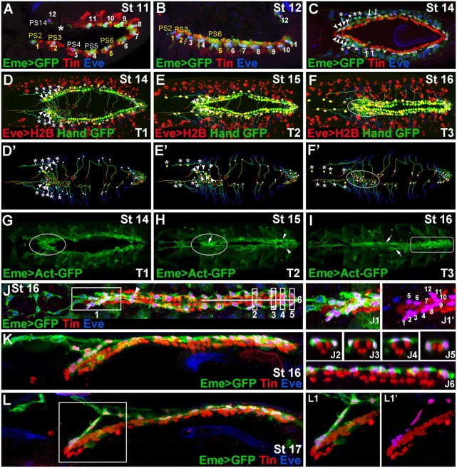
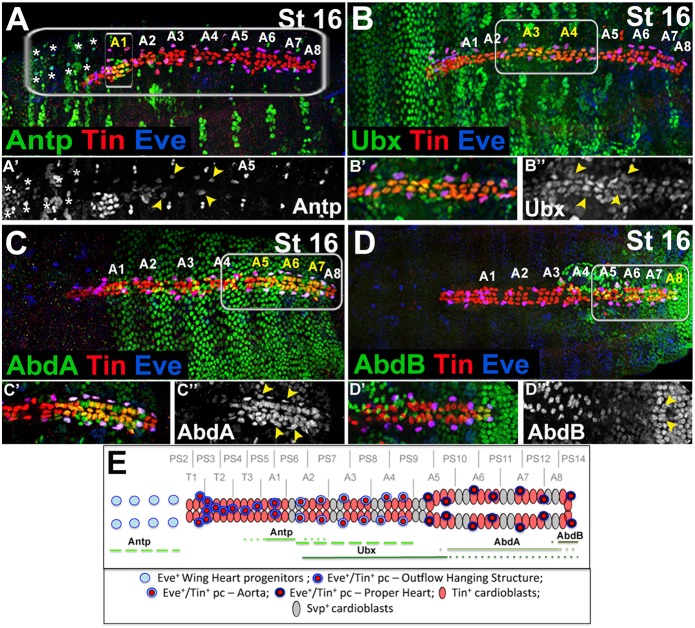
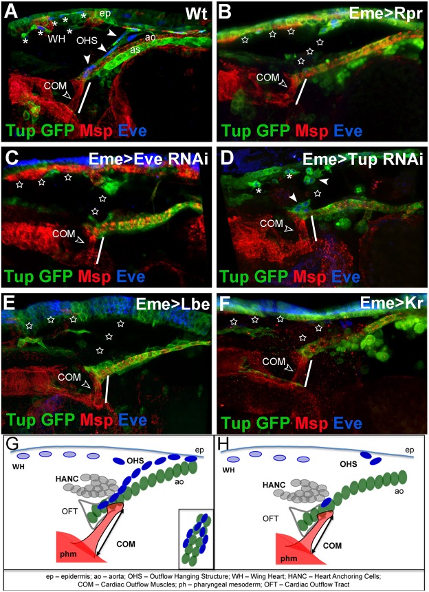
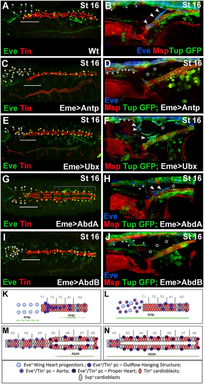
The Drosophila heart, composed of discrete subsets of cardioblasts and pericardial cells, undergoes Hox-triggered anterior-posterior morphogenesis, leading to a functional subdivision into heart proper and aorta, with its most anterior part forming a funnel-shaped cardiac outflow. Cardioblasts differentiate into Tin-positive 'working myocytes' and Svp-expressing ostial cells. However, developmental fates and functions of heart-associated pericardial cells remain elusive. Here, we show that the pericardial cells that express the transcription factor Even Skipped adopt distinct fates along the anterior-posterior axis. Among them, the most anterior Antp-Ubx-AbdA-negative cells form a novel cardiac outflow component we call the outflow hanging structure, whereas the Antp-expressing cells differentiate into wing heart precursors. Interestingly, Hox gene expression in the Even Skipped-positive cells not only underlies their antero-posterior diversification, but also influences heart morphogenesis in a non-cell-autonomous way. In brief, we identify a new cardiac outflow component derived from a subset of Even Skipped-expressing cells that stabilises the anterior heart tip, and demonstrate non-cell-autonomous effects of Hox gene expression in the Even Skipped-positive cells on heart morphogenesis.

Keywords: Drosophila; Heart; Hox genes; Pericardial cell.


Conflict of interest statement

Competing interestsThe authors declare no competing or financial interests.

Figures

Fig. 1.
Fig. 1.
Diversity of EPCs and formation of the OHS. (A,B) Lateral views from Eme>mcd8GFP stage 11 (A) and 12 (B) embryos showing 12 EPC clusters in dorsal mesoderm. The parasegments (PSs) from which OHS EPCs originate are coloured yellow. Asterisk in A indicates missing EPCs in PS13. (C) Dorsal view of a stage 14 embryo. Arrowheads indicate the OHS EPCs from PS2 and PS3; asterisks indicate detaching WH EPCs. The OHS EPCs from PS6 are indicated by arrows. (D-F′) Dorsal views of stage 14-16 living Hand-nlsGFP;Eme>H2B embryos showing cell tracking (lines) of migrating EPCs (white or yellow dots). (G-I) Dorsal views of Eme>lifeAct-GFP stage 14-16 living embryos. Leading edge activity of the migrating OHS EPCs is increased (G, circled). As the heart closure proceeds (H) the OHS EPCs become aligned (circled) with the apparent actin cable (arrow), whereas the heart proper EPCs (arrowheads) form a network of interconnected cells. At stage 16 (I), the actin cables (arrows) link the aorta EPCs, whereas the heart proper-associated EPCs (rectangle) display a patched actin enrichment. (J) Dorsal view of a stage 16 Eme>mcd8GFP embryo. (J1,J1ʹ) The OHS EPCs appear highly compacted and located dorsally above the heart tip. The EPCs associated with the aorta, except the first pair (arrowhead), lie more laterally and are interspaced according to the segmental register. The heart proper EPCs are located above or lateral to the cardioblasts (J2-J6), and appear as a dorsally connected network of cells. (K,L) Lateral views of stage 16 (K) and 17 (L) embryos stained as above and showing the arrangement of the heart-associated EPCs, which are interconnected. The OHS EPCs (L1,L1′) make a link between the bent anterior aorta and the epidermis.
Fig. 2.
Fig. 2.
Expression pattern of Hox proteins in the EPCs. (A-D″) Dorsal views of stage 16 embryos showing expression of Hox proteins. (A′-D″) Higher magnification views corresponding to areas framed by rectangles in A-D. (A,A′) Antp expression in the cardiac cells is confined to the A1 segment, including the A1 EPCs. There is also prominent expression of Antp in the WH EPCs (asterisks) and in EPCs from A2-A4 (yellow arrowheads). (B-B″) Ubx labels cardioblasts and pericardial cells, including EPCs (yellow arrowheads) from A3 and A4, and also cardiac cells from A2 and A5. Faint Ubx expression is seen in the heart proper. (C) High AbdA expression is detected in the cardioblasts and pericardial cells, including the EPCs (yellow arrowheads), from A6, A7 and the posterior part of A5. (D) AbdB expression is restricted to the most-posterior A8 cardioblasts and the pericardial cells, including the last pair of EPCs (yellow arrowheads). (E) Hox protein expression in the heart, including the EPCs. Segmental and parasegmental registers are depicted. Solid lines indicate the extent of Hox expression in the EPCs and cardioblasts. Dotted lines indicate the extent of Hox expression in cardioblasts. Dashed green lines indicate extended Antp expression in EPCs. No Hox expression could be detected in OHS EPCs from PS2 and PS3.
Fig. 3.
Fig. 3.
The OHS is required for cardiac outflow positioning. (A-F) Lateral views of the cardiac outflow region from Tup-GFP late stage 16 embryos. Tup-GFP reveals EPCs, aorta (ao), amnioserosa (as) and epidermis (ep); Msp300 stains COMs (open arrowhead) and cardiac cells; and Eve labels EPCs. (A) Wild-type embryo: the OHS EPCs are indicated by arrowheads and WH EPCs by asterisks. (B) Eme>Rpr embryo in which EPCs were ablated (stars). The COMs appear shorter (white line) than in wild type (compare A with B). (C) EPC-specific eveRNAi results in partial loss of OHS and WH EPCs (stars), and affects COMs length (white line). (D) EPC-targeted tupRNAi results in partial loss of EPCs. The COMs appear shorter (white line). (E,F) The eme-driven misexpression of Lbe (E) or Kr (F) represses eve, leading to affected specification of OHS and WH EPCs, and shortening of COMs (white lines). (G) Cellular components of cardiac outflow, including OHS. The OHS EPCs (dark blue) connect the tip of the anterior aorta (green) to the epidermis and, like COMs, are attached on both sides of the cardiac outflow. (H) Affected positioning of the cardiac outflow associated with COM shortening in the contexts where the OHS is lost.
Fig. 4.
Fig. 4.
Hox genes control EPCs diversity. (A,C,E,G,I) Dorsal views of late stage 16 embryos stained with Tin and Eve. (B,D,F,H,J) Lateral views of the cardiac outflow region from late stage 16 Tup-GFP embryos stained for GFP, Eve and Msp300. (A,B) In the wild-type embryos, the OHS EPCs are associated with the anterior aorta (A, above the line; B, arrowheads) and the WH EPCs are located anteriorly to the heart (asterisks). (C,D) In the Eme>Antp context, an increased number of EPCs adopt the WH fate (C,D, asterisks) and the OHS EPCs are almost absent (C, above the line; D, stars). (E,F) Eme-targeted overexpression of Ubx disrupts OHS morphogenesis (F, star) without shifting the identity of OHS EPCs to WH fate (E, above the line). Few OHS EPCs (arrowheads) are located more anteriorly compared with wild type (A). Asterisks indicate WH cells. The heart proper appears thinner. (G,H) In eme>AbdA embryos, the WH EPCs (asterisks) migrate more slowly, the string of OHS EPCs is broken (F, star) and the aorta is enlarged (G, bracket). (I,J) The Eme-driven expression of AbdB leads to a reduced EPC number. The OHS is missing (J, stars) and a few persisting EPCs adopt the WH fate (I, asterisks). In all EPC-targeted Hox misexpression experiments, the specification of the OHS EPCs is affected and the WH EPCs do not lose Tin expression. (K) Anterior aorta and WH progenitors in wild-type context. Solid and broken lines indicate Antp expression. (L) The shift in EPC identity to WH fate in the Eme-targeted Antp overexpression context. (M) The heart proper and posterior aorta organization with marked AbdA expression (solid line). (N) The extended heart proper phenotype observed in embryos with Eme-driven AbdA overexpression.

Similar articles

Cited by

References

    1. Alvarez A. D., Shi W., Wilson B. A. and Skeath J. B. (2003). pannier and pointedP2 act sequentially to regulate Drosophila heart development. Development 130, 3015-3026. 10.1242/dev.00488 - DOI - PubMed
    1. Bodmer R. and Frasch M. (2010). Development and aging of the Drosophila heart. In Heart Development and Regeneration (ed. Rosenthal N. and Harvey R. P.), pp. 47-86. Waltham, MA, USA: Academic Press.
    1. Brade T., Pane L. S., Moretti A., Chien K. R. and Laugwitz K.-L. (2013). Embryonic heart progenitors and cardiogenesis. Cold Spring Harb. Perspect. Med. 3, a013847 10.1101/cshperspect.a013847 - DOI - PMC - PubMed
    1. Brand A. H. and Perrimon N. (1993). Targeted gene expression as a means of altering cell fates and generating dominant phenotypes. Development 118, 401-415. - PubMed
    1. Carmena A., Gisselbrecht S., Harrison J., Jimenez F. and Michelson A. M. (1998a). Combinatorial signaling codes for the progressive determination of cell fates in the Drosophila embryonic mesoderm. Genes Dev. 12, 3910-3922. 10.1101/gad.12.24.3910 - DOI - PMC - PubMed

Publication types

MeSH terms