. 2017 Dec 15;7(1):17629.
doi: 10.1038/s41598-017-17772-6.

Increasing the permeability of Escherichia coli using MAC13243

Affiliations

Increasing the permeability of Escherichia coli using MAC13243

Claudio Muheim et al. Sci Rep. .

Abstract

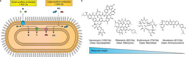
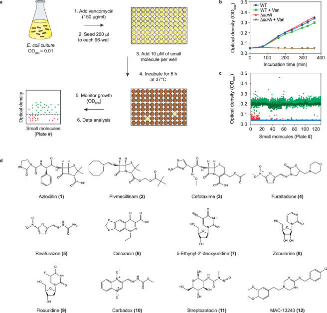
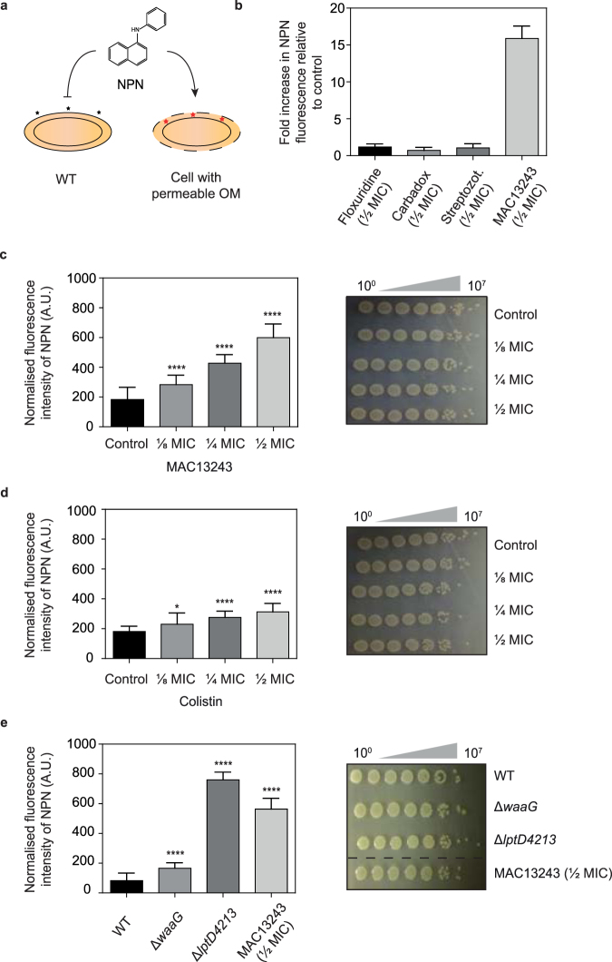
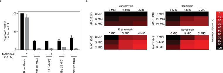
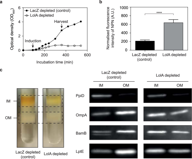
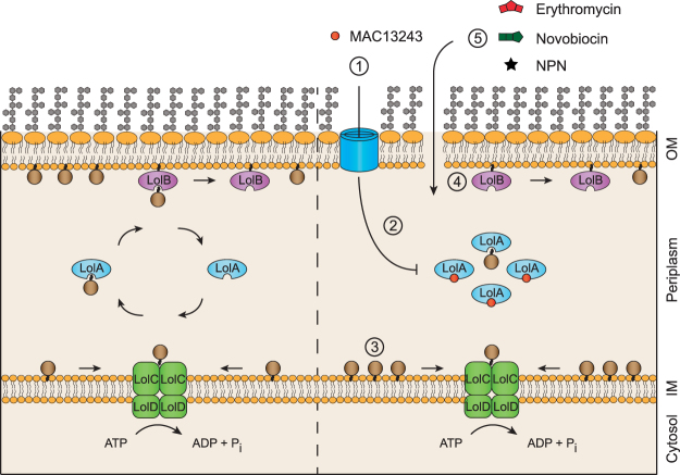
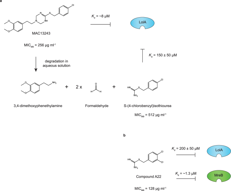
The outer membrane of gram-negative bacteria is a permeability barrier that prevents the efficient uptake of molecules with large scaffolds. As a consequence, a number of antibiotic classes are ineffective against gram-negative strains. Herein we carried out a high throughput screen for small molecules that make the outer membrane of Escherichia coli more permeable. We identified MAC13243, an inhibitor of the periplasmic chaperone LolA that traffics lipoproteins from the inner to the outer membrane. We observed that cells were (1) more permeable to the fluorescent probe 1-N-phenylnapthylamine, and (2) more susceptible to large-scaffold antibiotics when sub-inhibitory concentrations of MAC13243 were used. To exclude the possibility that the permeability was caused by an off-target effect, we genetically reconstructed the MAC13243-phenotype by depleting LolA levels using the CRISPRi system.


Conflict of interest statement

The authors declare that they have no competing interests.

Figures

Figure 1
Figure 1
Schematic illustration of antibiotic uptake in E. coli. (a) Antibiotics less than 600 Da (herein called small-scaffold antibiotics) can generally permeate through non-specific outer membrane porins and gain access to the periplasm. Antibiotics larger than 600 Da (herein called large-scaffold antibiotics) exceed the size exclusion limit of outer membrane porins. These antibiotics can presumably diffuse through the outer membrane but the process is inefficient. Once in the periplasm both small- and large-scaffold antibiotics can, in principle, diffuse across the inner membrane or can be inadvertently taken up by membrane embedded transporters. (b) Chemical structures of four large-scaffold antibiotics from different classes.
Figure 2
Figure 2
A high-throughput screen to identify small molecules that make E. coli more permeable. (a) An aliquot of E. coli MC4100 was seeded into individual wells of a 96-well microtiter plate in the presence of a sub MIC of vancomycin and small molecules. The plates were incubated for 5 h at 37 °C and cell growth (OD600) was compared to a DMSO control. (b) Growth curves of WT strain and a strain lacking the periplasmic chaperone SurA (ΔsurA) in the presence or absence of 150 μg mL−1 vancomycin (~1/3 MIC). The experiment indicates that growth of the WT strain is unaffected by a sub-lethal concentration of vancomycin, but growth of the ΔsurA is severely compromised. (c) Optical density readings of WT cells grown in the presence of 150 μg mL−1 vancomycin (~1/3 MIC) and 10 μM of each small molecule from the LCBKI library. 124 small molecules (red dots) inhibited growth more than 30% compared to the growth control (dark green dots). Sterility controls are illustrated by blue dots and small molecules that did not inhibit cell growth by more than 30% are illustrated as light green dots. (d) Chemical structures of 12 small molecules that inhibited cell growth in a vancomycin- and dose-dependent manner. 1–6 were from antibiotic classes that were previously known to function synergistically with vancomycin in gram-negative bacteria, and this category served as positive controls for the screen. 7–12 were not previously known to work in combination with vancomycin and they were chosen for follow up experiments.
Figure 3
Figure 3
A sub-lethal concentration of MAC13243 makes the outer membrane of E. coli more permeable. (a) The NPN dye can be used to monitor the integrity of the outer membrane. NPN is excluded from WT cells but penetrates into cells with a compromised outer membrane where it binds to the phospholipid layer, resulting in prominent fluorescence. (b) E. coli MC4100 grown in M9 media were exposed to different small molecules (½ MIC), and the permeability of the outer membrane was assessed by measuring the fluorescence of NPN. MICs were determined to be 1 μg mL−1 for carbadox, 256 μg mL−1 for streptozotocin, 0.002 μg mL−1 for floxuridine and 256 μg mL−1 for MAC13243. Fluorescence values were compared to cells treated with a solvent control. Note that we did not test all small molecules in the NPN uptake assay, but focussed on those that were readily available and that were representative of a class. For example, floxuridine (9) was deemed to be representative of the nucleoside analogues (7, 8). (c) E. coli MC4100 were grown in M9 media then exposed to different concentrations of MAC13234 (MIC = 256 μg mL−1) and NPN uptake was monitored (left panel). The increase in fluorescence was deemed to be due to increased permeability of the outer membrane, not cell lysis, since the amount of MAC13243 used did not affect cell viability (right panel). In these experiments cell aliquots were harvested after the NPN uptake assays, 10-fold serially diluted and spotted on LB agar. All data (mean ± SD) are from four experiments. ****p < 0.0001 (unpaired t-test.). (d) As for panel c except that E. coli MC4100 were exposed to different concentrations of colistin (MIC = 1 μg mL−1). (e) As for panel c except that the permeability of different E. coli strains was assessed.
Figure 4
Figure 4
MAC13243 makes E. coli more susceptible to some large-scaffold antibiotics. (a) E. coli MC4100 cells were exposed to 10 μM MAC13234, or large-scaffold antibiotics (½ MIC), or both. Cells were grown in LB for 18 h at 37 °C and cell growth was determined by measuring the optical density (OD600). MICs were determined to be 512 μg mL−1 for vancomycin, 16 μg mL−1 for rifampicin, 256 μg mL−1 for erythromycin and 64 μg mL−1 for novobiocin. (b) MAC13243 functions synergistically with some large-scaffold antibiotics. Heat plots showing growth inhibition of E. coli MC4100 in the presence of MAC13243 and vancomycin, rifampicin, erythromycin or novobiocin (in M9 media). Percentage of growth is illustrated with different colours where black represents 100% growth and red 0% growth. These data were used to calculate FICIs (see text for details). MICs were determined to be 256 μg mL−1 for MAC13243, 128 μg mL−1 for vancomycin, 8 μg mL−1 for rifampicin, 256 μg mL−1 for erythromycin and 1024 μg mL−1 for novobiocin.
Figure 5
Figure 5
Partial depletion of LolA increases the permeability of the outer membrane. (a) CRISPRi-mediated knockdown of LolA or LacZ in E. coli MC4100. Expression of dCas9 together with the respective lacZ sgRNA (control) or lolA sgRNA was induced at t = 90 min with 200 ng mL−1 aTC and growth was monitored by measuring optical density (OD600). (b) An aliquot of cells was taken 4 h after induction and permeability was monitored by the NPN uptake assay. All data (mean +/− S.D.) are from four experiments. ****p < 0.0001 (unpaired t-test.). (c) Depletion of LolA levels by CRISPRi affects the trafficking of both lipoproteins and ß-barrel proteins to the outer membrane. Inner and outer membrane fractions were purified from both LacZ-depleted cells (control) and LolA-depleted cells using a sucrose gradient. The proteins from each fraction were then separated by SDS-PAGE, transferred to a nitrocellulose membrane and probed with anti-sera to an inner membrane protein (PpiD), an outer membrane protein (OmpA), and two outer membrane lipoproteins (BamB and LptE). Full-length blots are shown in Supplementary Figure 4.
Figure 6
Figure 6
A model depicting how MAC13243 could affect the permeability of the outer membrane in E. coli. Lipoprotein precursors are synthesised on cytosolic ribosomes, then trafficked to the periplasm through either the Sec or Tat translocons. In the periplasm the N-terminal cysteine residue is acylated and then cleaved in successive reactions by Lgt, LspA and Lnt (not shown). Left panel, the mature lipoprotein is bound by the ABC transporter complex LolCDE then released to the periplasmic chaperone LolA. The LolA-lipoprotein complex is trafficked to the outer membrane where it binds to the LolB receptor and transfers the lipoprotein cargo. LolB then inserts the lipoprotein into the outer membrane. Note that some lipoproteins have a Lol avoidance signal and they are retained in the inner membrane. See for more details. Right panel, a sub-lethal concentration of MAC13243 (step 1) results in partial inhibition of LolA (step 2). This results in the partial retention of outer membrane lipoproteins at the inner membrane (step 3). Some of these mis-targeted lipoproteins are directly involved in outer membrane biogenesis, such as LolB (insertion of lipoproteins), BamB (insertion of ß-barrel proteins) and LptE (insertion of LPS molecules). Thus partial depletion of LolA can directly affect the biogenesis of the key components of the outer membrane, which weakens the membrane and results in increased permeability (step 4). The increased permeability in cells treated with MAC13243 can be exploited to increase the uptake of NPN and large-scaffold antibiotics (step 5).
Figure 7
Figure 7
MAC13243 is degraded in solution. (a) MAC13243 is hydrolysed into one molecule of 3,4-dimethoxyphenethylamine, two molecules of formaldehyde and one molecule of S-(4-chlorobenzyl)isothiourea. At neutral pH the t 1/2 is 13 h. Both MAC13243 and the degradation product S-(4-chlorobenzyl)isothiourea bind to LolA,. (b) An analogue of the degradation product, called A22 or S-(4-dichlorobenzyl)isothiourea, also binds LolA. Curiously this compound is a known inhibitor of the cytoskeletal protein MreB. Figure adapted from, with permission from the publisher.

Similar articles

Cited by

References

    1. Ruiz N, Kahne D, Silhavy TJ. Advances in understanding bacterial outer-membrane biogenesis. Nat. Rev. Microbiol. 2006;4:57–66. doi: 10.1038/nrmicro1322. - DOI - PubMed
    1. Silhavy TJ, Kahne D, Walker S. The bacterial cell envelope. Cold Spring Harb. Perspect. Biol. 2010;2:a000414. doi: 10.1101/cshperspect.a000414. - DOI - PMC - PubMed
    1. Nikaido H, Vaara M. Molecular basis of bacterial outer membrane permeability. Microbiol. Rev. 1985;49:1–32. - PMC - PubMed
    1. Nikaido H. Molecular basis of bacterial outer membrane permeability revisited. Microbiol. Mol. Biol. Rev. MMBR. 2003;67:593–656. doi: 10.1128/MMBR.67.4.593-656.2003. - DOI - PMC - PubMed
    1. Pagès J-M, James CE, Winterhalter M. The porin and the permeating antibiotic: a selective diffusion barrier in Gram-negative bacteria. Nat. Rev. Microbiol. 2008;6:893–903. doi: 10.1038/nrmicro1994. - DOI - PubMed

Publication types

MeSH terms