. 2017 Dec 15;7(1):17622.
doi: 10.1038/s41598-017-17914-w.

Generation of bioinspired structural colors via two-photon polymerization

Affiliations

Generation of bioinspired structural colors via two-photon polymerization

Gordon Zyla et al. Sci Rep. .

Abstract

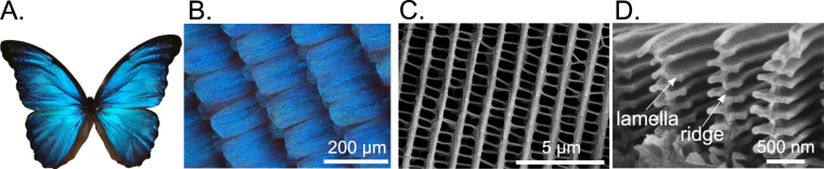
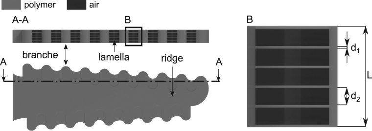
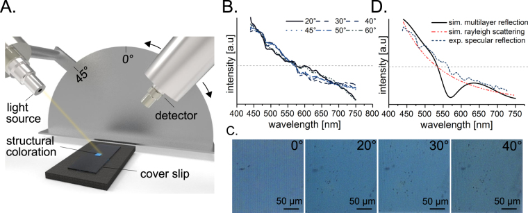
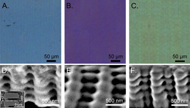
Colors of crystals, pigments, metals, salt solutions and bioluminescence occur in nature due to the optical properties of electrons in atoms and molecules. However, colors can also result from interference effects on nanostructures. In contrast to artificial coloration, which are caused by well-defined regular structures, the structural colors of living organisms are often more intense and almost angle-independent. In this paper, we report the successful manufacturing of a lamellar nanostructure that mimics the ridge shape of the Morpho butterfly using a 3d-direct laser writing technique. The viewing angle dependency of the color was analyzed via a spectrometer and the structure was visualized using a scanning electron microscope. The generated nano- and micro-structures and their optical properties were comparable to those observed in the Morpho butterfly.


Conflict of interest statement

The authors declare that they have no competing interests.

Figures

Figure 1
Figure 1
Structural coloration and wing composition of the M. didius. The blue coloration caused by the surface micro-/nano-structures is shown for a specimen of a M. didius in (A). The blue colored scales of the M. didius are presented in (B). A scanning electron microscope (SEM) image in (C) illustrates the arrangement of the M. didius ridges on a single wing scale. The cross section of the Morpho ridges and their Christmas tree shape contains alternating layers of air and natural material (cuticula) in the form of lamellas, as shown by the SEM image (D).
Figure 2
Figure 2
Computer-aided design of a single artificial photonic structure. The shape of the photonic structure on micron scale mimicking the artificial branches and ridge is shown in the top view. The selection drawing (A-A) illustrates the periodicity in the cross section of the microstructure. The periodicity consists of thin polymer conjunctions between each single branch, which mimic the Morpho lamellas. The artificial polymer lamellas are separated from each other through air cavities. The enlarged section (B) demonstrates the composition of one periodicity in detail. The thickness of one thin polymer film is defined with the value d 1. The size of one air is referenced with the value d 2. The parameter L denotes the total length of the periodicity.
Figure 3
Figure 3
Experimental 2PP set-up and detailed view of the polymerization inside a photosensitve material. The 2PP setup is illustrated schematically with an ultrashort pulse laser and a microscope objective (NA = 1.4) in (A). The principal of the 2PP manufacturing process inside a volume of polymer is demonstrated in (B).
Figure 4
Figure 4
Artificial blue coloration fabricated with photonic structures using 2PP. A section view of the polymerized array of three different laterally scaled photonic structures from the actual structure geometry (see Fig. 2) are shown in the SEM images (A)–(C). In comparison to sample (A), the variation in the lateral distances between each microstructure is illustrated in the SEM images for samples (B) and (C). The microscope images in (D)–(F) demonstrate the color formation results from those arrays.
Figure 5
Figure 5
Light scattering properties of photonic structures. The principal setup for spectral measurement of the coloration depending on the observer’s angle is illustrated in (A). A sample was illuminated at 45° and reflection spectra were measured at different angles. (B) The normalized to their area reflection spectra are shown at different observation angles. Upright light microscopy images (C) at different sample tilt angles were obtained using a microscope color camera. The simulation results of reflection spectra for the artificial photonic structure using two different approaches is illustrated in (D). A specular reflection for multilayer system consisting of 5 layers of the polymer separated by air layers is shown by solid line. Rayleigh scattering on multiple subwavelength centers is shown by dot/dash-line. Furthermore, the dash-line represents the specular reflection of the real photonic structure.
Figure 6
Figure 6
Mimicking the hierarchical surface micro-/nanostructures of the Morpho butterfly and coloration tuning by the variation of the phontonic structure’s size. The resulting coloration based on 2PP fabricated photonic structures are shown in (A)–(C). The associated SEM images of the microstructures are presented in (D)–(F). The oblique views of the microstructures demonstrates the hierarchical design containing air cavities and polymer lamellas. The total height of the microstructure was measured with white-light interferometry and is L = 1 μm. Since the number of periods is five, the total dimension of one air cavity and one artificial lamella for the resulting blue coloration (C) is 200 nm (d 1 ~ 50 nm, d 2 ~ 150 nm), which fits the air and cuticula dimensions inside the Christmas tree shape of a Morpho ridge. Whereas, the photonic microstrucutres of the purple coloration have decreased polymer lamellas d 1 ~ 40 nm and increased air cavities d 2 ~ 160 nm. The microstructures of the green coloration have the same amount regarding the dimensions of the polymer lamellas and air cavities (~100 nm).

Similar articles

Cited by

References

    1. Wickler W. Mimicry and the evolution of animal communication. Nature. 1965;208:519–521. doi: 10.1038/208519a0. - DOI
    1. Schultz TD, Fincke OM. Structural colours create a flashing cue for sexual recognition and male quality in a Neotropical giant damselfly. Funct. Ecol. 2009;23:724–732. doi: 10.1111/j.1365-2435.2009.01584.x. - DOI
    1. Arenas LM, Troscianko J, Stevens M. Color contrast and stability as key elements for effective warning signals. Front. Ecol. Evol. 2014;2:25. doi: 10.3389/fevo.2014.00025. - DOI
    1. Vukusic P, Sambles JR. Photonic structures in biology. Nature. 2003;424:852–855. doi: 10.1038/nature01941. - DOI - PubMed
    1. Ghiradella H. Light and color on the wing-structural colors in butterflies and moths. Appl. Opt. 1991;30:3492–3500. doi: 10.1364/AO.30.003492. - DOI - PubMed

LinkOut - more resources