. 2017 Dec 15;8(1):2146.
doi: 10.1038/s41467-017-01953-y.

Hepatocytic expression of human sodium-taurocholate cotransporting polypeptide enables hepatitis B virus infection of macaques

Affiliations

Hepatocytic expression of human sodium-taurocholate cotransporting polypeptide enables hepatitis B virus infection of macaques

Benjamin J Burwitz et al. Nat Commun. .

Abstract

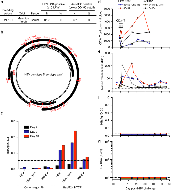
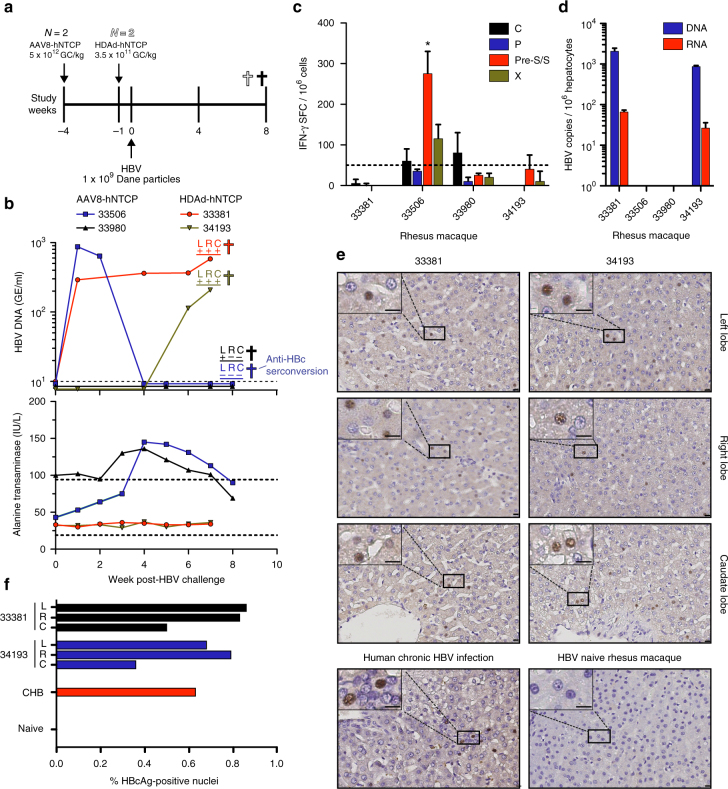
Hepatitis B virus (HBV) is a major global health concern, and the development of curative therapeutics is urgently needed. Such efforts are impeded by the lack of a physiologically relevant, pre-clinical animal model of HBV infection. Here, we report that expression of the HBV entry receptor, human sodium-taurocholate cotransporting polypeptide (hNTCP), on macaque primary hepatocytes facilitates HBV infection in vitro, where all replicative intermediates including covalently closed circular DNA (cccDNA) are present. Furthermore, viral vector-mediated expression of hNTCP on hepatocytes in vivo renders rhesus macaques permissive to HBV infection. These in vivo macaque HBV infections are characterized by longitudinal HBV DNA in serum, and detection of HBV DNA, RNA, and HBV core antigen (HBcAg) in hepatocytes. Together, these results show that expressing hNTCP on macaque hepatocytes renders them susceptible to HBV infection, thereby establishing a physiologically relevant model of HBV infection to study immune clearance and test therapeutic and curative approaches.


Conflict of interest statement

Oregon Health and Science University (OHSU), Dr. Sacha, and Dr. Burwitz have a financial interest in Vir Biotechnology, Inc., a company that may have a financial interest in the results of this research and technology. This potential individual and institutional conflict of interest has been reviewed and managed by OHSU. The remaining authors declare no competing financial interests.

Figures

Fig. 1
Fig. 1
Mauritian-origin HBV (mcHBV) does not infect MCM in vitro or in vivo. a Twenty-seven MCM caught on the island of Mauritius and housed at the ONPRC were screened for HBV DNA and anti-HBc. b Schematic of HBV proteome (black) showing amino acid differences for the recently described mcHBV. * = HBV sequence described by Pasek et al.. c Cynomolgus PH and HepG2-hNTCP cells were infected with HBV, HBV P68S, and mcHBV (MOI = 100) and infection monitored by HBsAg ELISA. Dotted lines represent limit of detection. dg Four MCM were challenged with either HBV P68S or mcHBV (1 × 109 virions). Each condition represents a single-biological sample (N = 1). Figure is representative data of two separate experiments. d One MCM from each group received CD3-immunotoxin (CD3-IT) and absolute CD3+ T-cell frequencies were monitored over time. HBV infection was monitored longitudinally by measurements of e ALT (dotted lines represent normal ALT reference range in the ONPRC colony), f HBsAg (dotted lines represent limit of detection), and g HBV DNA (dotted lines represent limit of detection)
Fig. 2
Fig. 2
In vitro HBV infection of rhesus macaque PH. a Predicted schematic of NTCP showing amino acid differences between human and macaque NTCP. Differences in the sequences were labeled with lighter red for amino acid exchanges with similar physiochemical properties and darker red for exchanges with different physiochemical properties. Gray box represents cellular membrane. N-linked glycosylation sites represented by black brackets. macNTCP = macaque NTCP. b Rhesus macaque PH were transduced with either HDAd-hNTCP (MOI = 2) or AAV-hNTCP (MOI = 1 × 104) and stained 3 days later with Myrcludex B-atto488. c Rhesus macaque and baboon PH were transduced with either HDAd-hNTCP (MOI = 2) or AAV-hNTCP (MOI = 1 × 104) and infected with HBV (MOI = 100) 3 days later. Productive infection was monitored by quantification of HBsAg and HBeAg in the supernatant by ELISA. Each condition represents a single-biological sample (N = 1). Figure is representative data of two separate experiments. d HBV DNA qPCR on the same supernatants shown in c. Each condition represents a single-biological sample (N = 1). e Total intracellular DNA from 1 × 106 rhesus macaque PH and HepG2-hNTCP cells was used in a cccDNA-specific qPCR. Rhesus macaque PH transduced with AAV-hNTCP (MOI = 1 × 104) and infected with HBV (MOI = 100) 3 days later showed formation of cccDNA, while the non-transduced, HBV challenged PH did not. Bars represent standard error of measurement from two qPCR replicates. f Southern blot shows presence of cccDNA in rhesus macaque PH transduced with AAV-hNTCP (MOI = 1 × 104) and infected with HBV (MOI = 100). DNA was purified after Hirt extraction to remove protein-bound DNA forms. SM = size marker; T5 Exo = T5 exonuclease; PF-rcDNA = polymerase-free relaxed circular DNA; PF-dlDNA = polymerase-free duplex linear DNA. Figure is representative data of two separate experiments. g Neonate rhesus macaque PH were transduced with AAV-hNTCP (MOI = 1 × 104 or 5 × 102) and infected with HBV (MOI = 100) 3 days later. HBV infection was then monitored longitudinally by HBsAg ELISA. Each condition represents a single-biological sample (N = 1)
Fig. 3
Fig. 3
In vivo HBV infection of rhesus macaques. a Timeline showing administration of viral vectors encoding hNTCP, HBV challenge, and necropsy of rhesus macaques. GC = genome copies. b Longitudinal monitoring of HBV infection by HBV DNA (dotted line represents assay limit of detection) and alanine transaminase concentrations (dotted lines represent normal alanine transaminase reference range in ONPRC colony) in the serum. c IFN-g ELISpot measurements of liver-resident T-cell responses against HBV C, P, S, and X. * = Response met statistical significance, defined as mean number of spot forming cells (SFCs) of triplicate sample wells exceeding background (no stim) plus two standard deviations in a homoscedastic t-test. Dotted line represents assay limit of detection. d Levels of HBV DNA and RNA as assessed by qPCR and qRT-PCR, respectively, in rhesus macaque PH isolated from the liver via collagenase media perfusion. e HBcAg immunohistochemistry from HBV-infected rhesus macaque liver lobes, HBV naive rhesus macaque liver, and chronically HBV-infected human liver. Size bars indicate distance of 10 μm. f Quantification of HBcAg immunohistochemistry in e showing average frequency of HBcAg-positive nuclei (include parenchymal and non-parenchymal cells) from indicated liver lobes. Human CHB and naive rhesus macaque samples represent a single-biological sample (N = 1). CHB = Human chronic HBV infection, L = left liver lobe, R = right liver lobe, C = caudate liver lobe

Similar articles

Cited by

References

    1. Franco E, et al. Hepatitis B: epidemiology and prevention in developing countries. World J. Hepatol. 2012;4:74–80. doi: 10.4254/wjh.v4.i3.74. - DOI - PMC - PubMed
    1. Ely A, Arbuthnot P. Differing prospects for the future of using gene therapy to treat infections with hepatitis B virus and hepatitis C virus. Discov. Med. 2015;20:137–143. - PubMed
    1. Andersson, K. L. & Chung, R. T. in Monitoring during and after antiviral therapy for hepatitis B. Vol. 49 (ed. Hoofnagle, J. H.) 166–S173 (Wiley Subscription Services, Inc., A Wiley Company, 2009).
    1. Urban S, Breiner KM, Fehler F, Klingmüller U, Schaller H. Avian hepatitis B virus infection is initiated by the interaction of a distinct pre-S subdomain with the cellular receptor gp180. J. Virol. 1998;72:8089–8097. - PMC - PubMed
    1. Fukuda R, Fukumoto S, Shimada Y. A sequential study of viral DNA in serum in experimental transmission of duck hepatitis B virus. J. Med. Virol. 1987;21:311–320. doi: 10.1002/jmv.1890210403. - DOI - PubMed

Publication types

MeSH terms