. 2018 Jan;15(1):216-232.
doi: 10.1007/s13311-017-0591-2.

iTRAQ-Based Quantitative Proteomics Reveals the New Evidence Base for Traumatic Brain Injury Treated with Targeted Temperature Management

Affiliations

iTRAQ-Based Quantitative Proteomics Reveals the New Evidence Base for Traumatic Brain Injury Treated with Targeted Temperature Management

Shi-Xiang Cheng et al. Neurotherapeutics. 2018 Jan.

Abstract

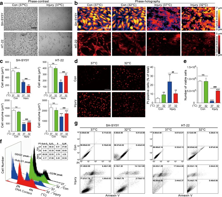
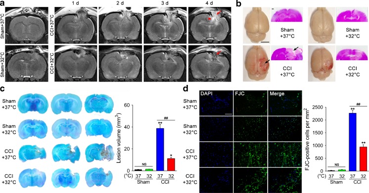
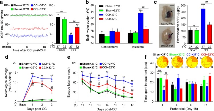
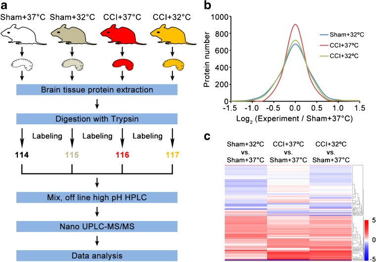
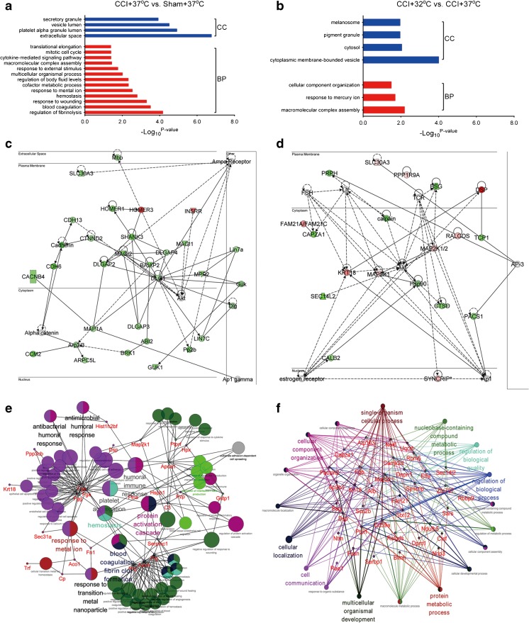
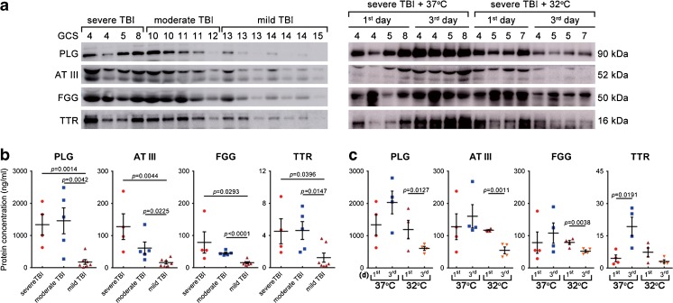
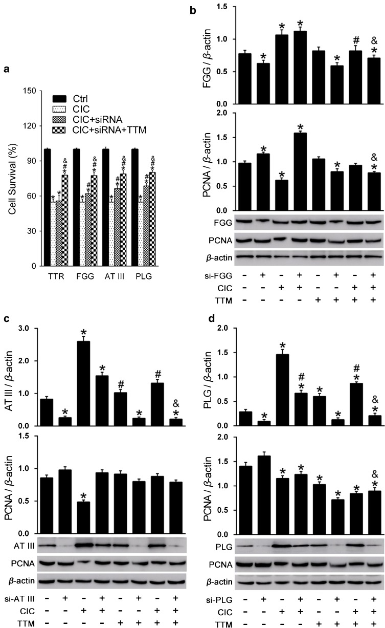
This study aimed to investigate the effects of targeted temperature management (TTM) modulation on traumatic brain injury (TBI) and the involved mechanisms using quantitative proteomics technology. SH-SY5Y and HT-22 cells were subjected to moderate stretch injury using the cell injury controller (CIC), followed by incubation at TTM (mild hypothermia, 32°C), or normothermia (37°C). The real-time morphological changes, cell cycle phase distribution, death, and cell viability were evaluated. Moderate TBI was produced by the controlled cortical impactor (CCI), and the effects of TTM on the neurological damage, neurodegeneration, cerebrovascular histopathology, and behavioral outcome were determined in vivo. Results showed that TTM treatment prevented TBI-induced neuronal necrosis in the brain, achieved a substantial reduction in neuronal death both in vitro and in vivo, reduced cortical lesion volume and neuronal loss, attenuated cerebrovascular histopathological damage, brain edema, and improved behavioral outcome. Using an iTRAQ proteomics approach, proteins that were significantly associated with TTM in experimental TBI were identified. Importantly, changes in four candidate molecules (plasminogen [PLG], antithrombin III [AT III], fibrinogen gamma chain [FGG], transthyretin [TTR]) were verified using TBI rat brain tissues and TBI human cerebrospinal fluid (CSF) samples. This study is one of the first to investigate the neuroprotective effects of TTM on the proteome of human and experimental models of TBI, providing an overall landscape of the TBI brain proteome and a scientific foundation for further assessment of candidate molecules associated with TTM for the promotion of reparative strategies post-TBI.

Keywords: Cerebrospinal fluid; Isobaric tags for relative and absolute quantitation; Proteomics; Targeted temperature management; Traumatic brain injury.


Figures

Fig. 1
Fig. 1
Effects of targeted temperature management (TTM) on SH-SY5Y and HT-22 cells with traumatic brain injury-like damage. SH-SY5Y and HT-22 cells were either uninjured exposed to TTM (32°C) or normothermia (37°C), or subjected to stretch injury and harvested either 32°C or 37°C treatment for 6 h. The morphological changes were detected by (a) phase-contrast microscopy and (b) phase-holographic microscopy. (c) The mean cell area and volume were measured by digital holographic microscopy assay. (d) SH-SY5Y cell death was analyzed by propidium iodine (PI) labeling and counts were performed on PI-positive cells. (e) Quantification of SH-SY5Y cell viability was then measured using 0.4% trypan blue with the automated Countess system. (f) Representative flow cytometry analysis of SH-SY5Y cell-cycle distribution, expressed as the percentage of cells at sub-G1, G0/G1, S, and G2/M phase. (g) Necrosis and apoptosis were measured with Annexin V/PI labeling by flow cytometry. *p < 0.05, **p < 0.01, ***p < 0.001 vs uninjured control (37°C); ## p < 0.01 and ### p < 0.001 vs injured cells (37°C). Error bars represent SD (n = 3 independent experiments). NS indicates p > 0.05
Fig. 2
Fig. 2
Effects of targeted temperature management (TTM) on neurological damage and neurodegeneration after traumatic brain injury (TBI). (a) Representative T2-weighted magnetic resonance images at 1–4 days post-TBI. TTM reduces lesion size (arrow) and trauma-associated hydrocephalus (arrowhead) (n = 6/group). (b) Cortical contusion impact (CCI)-induced TBI created a focal lesion (arrow) in the right hemisphere of the rat brain (scale bar = 5 mm) at 24 h post-TBI. Representative images of hematoxylin and eosin-stained sections, highlighting considerable sparing of brain tissue in CCI + 32°C rats (scale bar = 500 μm). (c) At 24 h postinjury, brains were sectioned and stained with cresyl violet. Contralateral (left hemisphere) and ipsilateral (right hemisphere) substructure volumes were calculated in multiple sections per rat. Representative images from 3 consecutive sections per rat. Quantification of lesion volume was then assessed by subtracting the ipsilateral volume from the contralateral volume. (d) Assessment of neurodegeneration by Fluoro-Jade C (FJC) staining. Neurodegeneration observed in the pericontusional area 24 h after CCI was attenuated by TTM (scale bar = 40 μm). *p < 0.05 and **p < 0.01 vs sham + 37°C; ## p < 0.01 vs CCI + 37°C. Error bars represent SD (n = 3/group). DAPI = 4’,6-diamidino-2-phenylindole. NS indicates p > 0.05
Fig. 3
Fig. 3
Effects of targeted temperature management (TTM) on cerebrovascular histopathology and behavioral outcome post-traumatic brain injury (TBI). (a) TTM increases regional cerebral blood flow (rCBF) 24 h after TBI. Representative original recording of ipsilateral rCBF for 10 min. Values are expressed as mean ± SD (n = 6/group). (b) Graph showing increased brain water content (determined by dry:wet ratio) in the traumatized hemisphere 24 h post-TBI, whereas TTM decreases the accumulation of water in the tissue (n = 3/group). (c) Blood–brain barrier integrity was measured by assessing the extravasation of Evans blue (EB) dye. The extravasation of EB dye in brain samples from cortical contusion impact (CCI) + 37°C or 32°C rats is shown. (d) Modified neurological severity (mNSS) score was assigned 1, 2, and 3 days post-TBI (n = 20/group). (e) The Morris water maze test was used to detect the spatial learning and memory for 7 consecutive days, starting on post-injury day (PID) 11 after TBI (n = 20/group). TTM significantly shortened escape latency during PID 12–17. (f) Representative path tracings in each quadrant during the probe trial on PID 18 (T = target quadrant; R = right quadrant; O = opposite quadrant; L = left quadrant). **p < 0.01 and ***p < 0.001 vs sham (37°C); # p < 0.05 and ## p < 0.01 vs CCI (37°C). Error bars represent SD. NS indicates p > 0.05
Fig. 4
Fig. 4
Quantitative proteomics comparison of the cortical contusion impact (CCI) rats treated with targeted temperature management using the iTRAQ approach. (a) Workflow of quantitative proteomics by iTRAQ. (b) Histograms of log2 ratio distributions of quantified proteins: sham + 32°C (n = 1526), cortical contusion impact (CCI) + 37°C (n = 1523), and CCI + 32°C (n = 1529) vs sham + 37°C, respectively. (c) Heat map of differentially expressed proteins in sham + 32°C (n = 153), CCI + 37°C (n = 73), and CCI + 32°C (n = 130) vs sham + 37°C, respectively. HPLC = high-performance liquid chromatography; UPLC = ultra performance liquid chromatography; MS/MS = tandem mass spectrometry
Fig. 5
Fig. 5
Bioinformatics analysis for the differentially expressed proteins in the cortical contusion impact (CCI) rats treated with targeted temperature management. Diagram showing the biological process (BP) and cellular component (CC) of differentially expressed proteins of (a) CCI + 37°C vs sham + 37°C and (b) CCI + 32°C vs CCI + 37°C using DAVID analysis. Network of (c) CCI + 37°C vs sham + 37°C and (d) CCI + 32°C vs CCI + 37°C clustered by Ingenuity Pathway Analysis. Protein–protein interaction was generated by Cytoscape between (e) CCI + 37°C vs sham + 37°C and (f) CCI + 32°C vs CCI + 37°C
Fig. 6
Fig. 6
Plasminogen (PLG), antithrombin III (AT III), fibrinogen gamma chain (FGG), and transthyretin (TTR) levels reflected injury magnitude, and the effect of TTM post-traumatic brain injury (TBI). (a) The levels of PLG, AT III, FGG, and TTR in 28 cerebrospinal fluid (CSF) samples were evaluated by Western blot, including 4 patients with severe TBI (Glasgow Coma Scale [GCS] 3–8), 5 with moderate TBI (GCS 9–12), 7 with mild TBI (GCS 13–15) with standard therapy at 1 day after trauma (left panel), and 4 patients with severe TBI (the same patients with that in the left panel) followed by standard therapy (37°C) and 4 with severe TBI followed by TTM + standard therapy (32°C) at 1 and 3 days postinjury (right panel). (b) Temporal profile of CSF PLG, AT III, FGG, and TTR levels (ng/ml) among patients with severe (n = 4), moderate (n = 5), and mild TBI (n = 7) with standard therapy at 1 day after trauma, and (c) in patients with severe TBI receiving standard therapy and TTM + standard therapy at 1 and 3 days postinjury were measured by enzyme-linked immunosorbent assay. Error bars represent SD
Fig. 7
Fig. 7
Validation of the influence of plasminogen (PLG), antithrombin III (AT III), fibrinogen gamma chain (FGG), and transthyretin (TTR) on HT-22 cell viability by small interfering RNA (siRNA) treatment. (a) PLG, AT III, FGG, and TTR were knocked down in cells. Then targeted temperature management (TTM) was applied and CellTiter 96 AQueous One solution Cell Proliferation Assay was used to measure cell viability. (b) Effects of FGG knockdown and TTM on the expression of FGG and proliferating cell nuclear antigen (PCNA). (c) Effects of AT III knockdown and TTM on the expression of AT III and PCNA. (d) Effects of PLG knock down and TTM on the expression of PLG and PCNA. *p < 0.05 vs the control group; # p < 0.05 vs the traumatic brain injury (TBI) group; & p < 0.05 vs the TBI + siRNA knockdown group. CIC = cell injury controller

Similar articles

Cited by

References

    1. Manley GT, Maas AI. Traumatic brain injury: an international knowledge-based approach. JAMA. 2013;310:473–474. doi: 10.1001/jama.2013.169158. - DOI - PubMed
    1. Jiang JY. Head trauma in China. Injury. 2013;44:1453–1457. doi: 10.1016/j.injury.2012.08.045. - DOI - PubMed
    1. Corrigan JD, Selassie AW, Orman JA. The epidemiology of traumatic brain injury. J Head Trauma Rehabil. 2010;25:72–80. doi: 10.1097/HTR.0b013e3181ccc8b4. - DOI - PubMed
    1. Nunnally ME, Jaeschke R, Bellingan GJ, et al. Targeted temperature management in critical care: a report and recommendations from five professional societies. Crit Care Med. 2011;39:1113–1125. doi: 10.1097/CCM.0b013e318206bab2. - DOI - PubMed
    1. Zhao CC, Wang CF, Li WP, et al. Mild hypothermia promotes pericontusion neuronal sprouting via suppressing suppressor of cytokine signaling 3 expression after moderate traumatic brain injury. J Neurotrauma. 2017;34:1636–1644. doi: 10.1089/neu.2016.4759. - DOI - PubMed

Publication types

MeSH terms