. 2018 Jan 25:280:109-116.
doi: 10.1016/j.cbi.2017.12.019. Epub 2017 Dec 13.

Effects of a novel microtubule-depolymerizer on pro-inflammatory signaling in RAW264.7 macrophages

Affiliations

Effects of a novel microtubule-depolymerizer on pro-inflammatory signaling in RAW264.7 macrophages

Samuel P Gilmore et al. Chem Biol Interact. .

Abstract

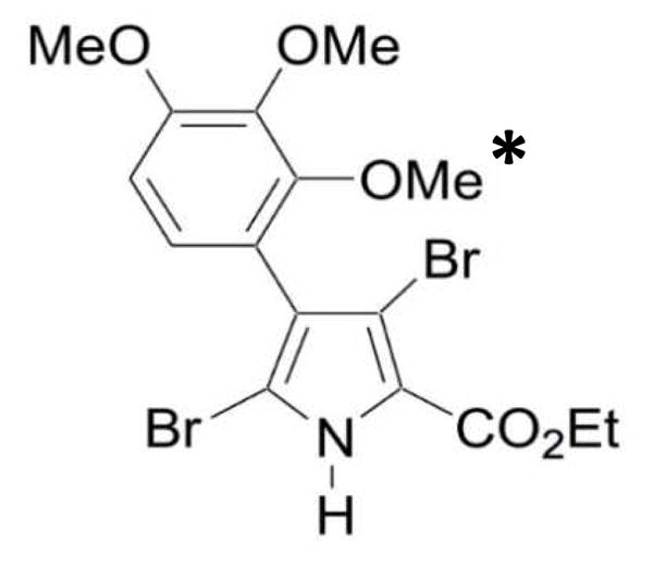
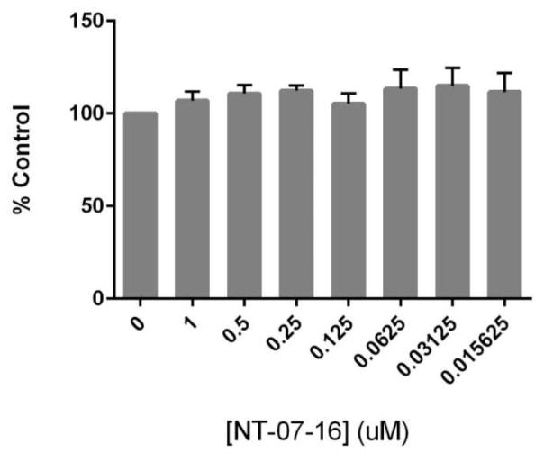
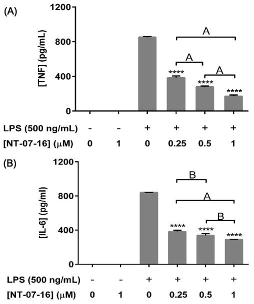
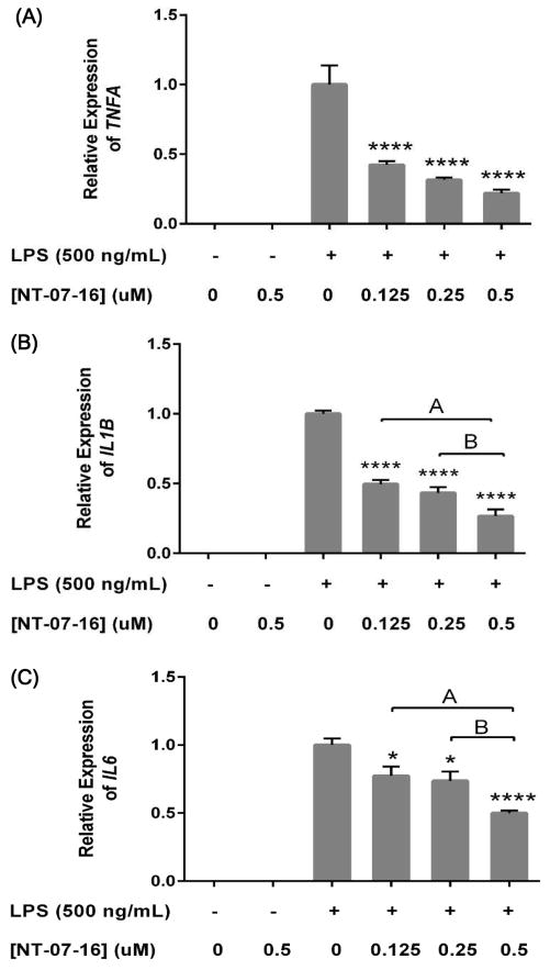
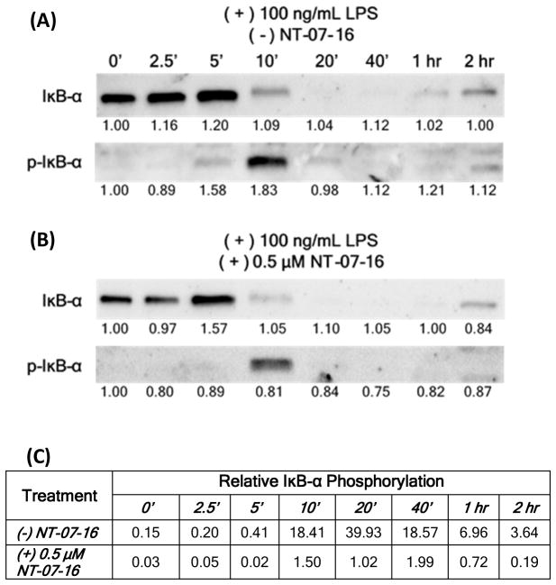
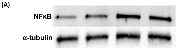
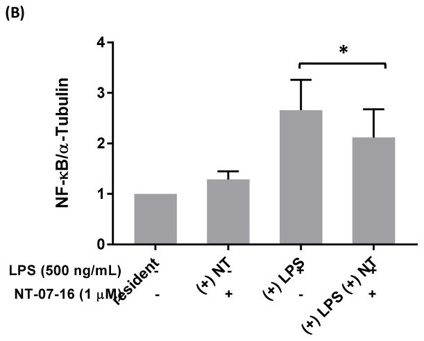
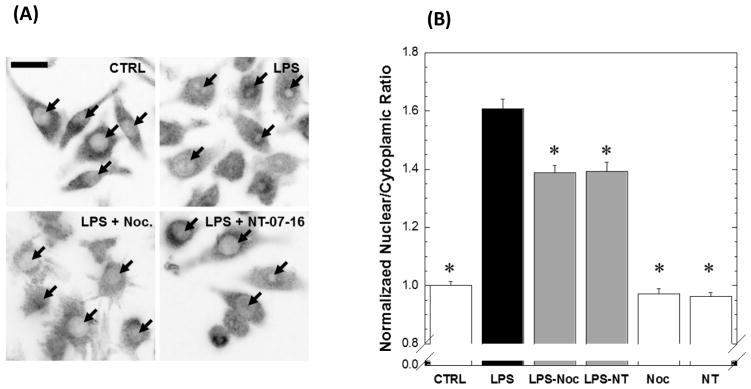
The Nuclear Factor-kappa B (NF-κB) pathway is vital for immune system regulation and pro-inflammatory signaling. Many inflammatory disorders and diseases, including cancer, are linked to dysregulation of NF-κB signaling. When macrophages recognize the presence of a pathogen, the signaling pathway is activated, resulting in the nuclear translocation of the transcription factor, NF-κB, to turn on pro-inflammatory genes. Here, we demonstrate the effects of a novel microtubule depolymerizer, NT-07-16, a polysubstituted pyrrole compound, on this process. Treatment with NT-07-16 decreased the production of pro-inflammatory cytokines in RAW264.7 mouse macrophages. It appears that the reduction in pro-inflammatory mediators produced by the macrophages after exposure to NT-07-16 may be due to activities upstream of the translocation of NF-κB into the nucleus. NF-κB translocation occurs after its inhibitory protein, IκB-α is phosphorylated which signals for its degradation releasing NF-κB so it is free to move into the nucleus. Previous studies from other laboratories indicate that these processes are associated with the microtubule network. Our results show that exposure to the microtubule-depolymerizer, NT-07-16 reduces the phosphorylation of IκB-α and also decreases the association of NF-κB with tubulin which may affect the ability of NF-κB to translocate into the nucleus. Therefore, the anti-inflammatory activity of NT-07-16 may be explained, at least in part, by alterations in these steps in the NF-κB signaling pathway leading to less NF-κB entering the nucleus and reducing the production of pro-inflammatory mediators by the activated macrophages.

Keywords: Inflammation; Macrophage; Microtubule; NF-κB; NT-07-16 pyrrole compound; Signaling.


Figures

Figure 1
Figure 1
Structure of NT-07-16. An additional methoxy group (marked with an *) was added to the 2-position on the phenyl ring of the parent compound, JG-03-14 (not shown), in order to enhance its ability to interact with tubulin.
Figure 2
Figure 2
Effect of NT-07-16 on RAW264.7 cell viability. Cells were exposed to varying concentrations of NT-07-16 for 20 hours. Viability was assessed using an MTT assay and is reported as a percentage of the control sample (macrophages not exposed to NT-07-16). Results are representative of three independent experiments and error bars indicate the standard error of the mean within each treatment group.
Figure 3
Figure 3
Effect of NT-07-16 on IL-6 and TNFα production by activated RAW264.7 macrophages. Cells were exposed to NT-07-16 for one hour prior to activation with LPS. Cells were incubated for 20 hours and then supernatants were collected. OptEIA™ ELISA Assay kits were used to quantify (A) TNF-α and (B) IL-6 released from the cells. Variation among treatments was determined by one-way ANOVA. The means of each sample were compared to the LPS control, **** p<0.0001; significant differences in expression were seen in independently treated samples, A= p<0.01, B= p<0.05 (Tukey-Kramer post-hoc test). Results are representative of three independent experiments and error bars indicate the standard error of the mean within each treatment group.
FIGURE 4
FIGURE 4
Effect of NT-07-16 on TNFA, IL1B, and IL6 expression in RAW264.7 macrophages. Cells were exposed to NT-07-16 for one hour prior to activation with LPS. Cells were incubated for 4 hours before RNA isolation. Relative expression of (A) TNFA, (B) IL1B, and (C) IL6 was measured using qPCR with β-actin as the reference gene. Variation among treatments was determined by one-way ANOVA. The means of each sample were compared to the LPS, * p<0.05, **** p<0.0001; significant differences in expression were also seen between independently treated samples, A= p<0.01, B= p<0.05 (Tukey-Kramer post-hoc test). Results are representative of three independent experiments and error bars indicate the standard error of the mean within each treatment group.
Figure 5
Figure 5
Effect of NT-07-16 on the phosphorylation of IκB-α over time. RAW264.7 macrophages were activated with LPS for the indicated length of time and cell lysates were prepared and analyzed by Western blotting using antibodies specific for IκB-α and p-IκB-α. Macrophages were either (A) not exposed to or (B) were exposed to 0.5 μM NT-07-16 for one hour prior to LPS activation. Normalization factors are below each blot. These values were determined by dividing the total adjusted protein volume (background subtracted) of each lane by the total adjusted protein volume of lane 1 on each blot. (C) Relative IκB-α phosphorylation was calculated by dividing the normalized densitometry values of p-IκB-α by the normalized densitometry values of IκB-α for each time period. Low values indicate little p-IκB-α compared to the unphosporylated form for each time period whereas high values indicate high levels of pIκB-α compared to the unphosporylated form. Results are representative of three independent experiments.
Figure 6
Figure 6
Effect of NT-07-16 on the association of NF-κB with tubulin in RAW264.7 macrophages. Cells were exposed to NT-07-16 for 1 hour prior to activation with LPS for 30 minutes. Total cell lysates were collected and immunoprecipitation was performed to isolate α-tubulin. The association of NF-κB with α-tubulin was analyzed by western blotting (A). Densitometry was performed to analyze the amount of NF-κB that co-immunoprecipitated with α-tubulin before and after exposure to NT-07-16 in control and LPS-activated macrophages (B). Data incorporate results from four independent experiments. * p<0.05 (paired t-test).
Figure 6
Figure 6
Effect of NT-07-16 on the association of NF-κB with tubulin in RAW264.7 macrophages. Cells were exposed to NT-07-16 for 1 hour prior to activation with LPS for 30 minutes. Total cell lysates were collected and immunoprecipitation was performed to isolate α-tubulin. The association of NF-κB with α-tubulin was analyzed by western blotting (A). Densitometry was performed to analyze the amount of NF-κB that co-immunoprecipitated with α-tubulin before and after exposure to NT-07-16 in control and LPS-activated macrophages (B). Data incorporate results from four independent experiments. * p<0.05 (paired t-test).
Figure 7
Figure 7
Pretreatment of RAW264.7 cells with microtubule depolymerizing compounds disrupts the typical microtubule networking phenotype. Qualitative image analysis demonstrates microtubule depolymerization of RAW264.7 cells treated with either nocodazole or NT-07-16. Arrows indicate the altered microtubule network in the nocodazole and NT-07-16 treated macrophages. Scale bar = 20 μm.
Figure 8
Figure 8
Effect of microtubule depolymerization on nuclear translocation of NFκB in RAW264.7 macrophages. (A) Intensity-reversed representative images of p65 staining in control RAW264.7 macrophages, or cells activated with 500 ng/ml of LPS, in the absence or presences of 1μM nocodazole, or 1μM NT-07-16. Arrows represent position of nuclei. Scale bar = 20μm. (B) Normalized mean nuclear localization ratios were calculated from in vitro immunofluorescence imaging of the p65 subunit of NF-κB. LPS activation resulted in an increase in nuclear NF-κB compared to cells that were not activated. Exposure to either nocodazole or NT-07-16 for 1 hour prior to LPS activation decreased the response (*Dunnett’s Test compared to LPS treatment, p < 0.0001). Error bars represent standard error of the mean. n >190 cells for each condition across five independent experiments.

References

    1. Antonelli A, Kushner I. It’s time to redefine inflammation. FASEB J. 2017;31:1787–1791. - PubMed
    1. Kondylis V, Kumari S, Vlantis K, Pasparakis M. The interplay of IKK, NF- B and RIPK1 signaling in the regulation of cell death, tissue homeostasis and inflammation. Immunol Rev. 2017;277:113–127. - PubMed
    1. Munn LL. Cancer and inflammation. WIREs Syst Biol Med. 2017 Mar-Apr; doi: 10.1002/wsbm.1370. - DOI - PMC - PubMed
    1. Gordon S. Pattern recognition receptors: doubling up for the innate immune response. Cell. 2002;111:927–930. - PubMed
    1. Komai K, Shichita T, Ito M, Kanamori M, Chikuma S. Role of scavenger receptors as damage-associated molecular pattern receptors in Toll-like receptor activation. Int Immunol. 2017;29:59–70. - PubMed

MeSH terms

LinkOut - more resources