. 2018 Mar 15:137:55-66.
doi: 10.1016/j.ymeth.2017.12.004. Epub 2017 Dec 14.

Structural dynamics of protein S1 on the 70S ribosome visualized by ensemble cryo-EM

Affiliations

Structural dynamics of protein S1 on the 70S ribosome visualized by ensemble cryo-EM

Anna B Loveland et al. Methods. .

Abstract

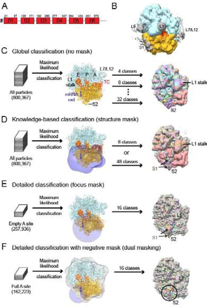
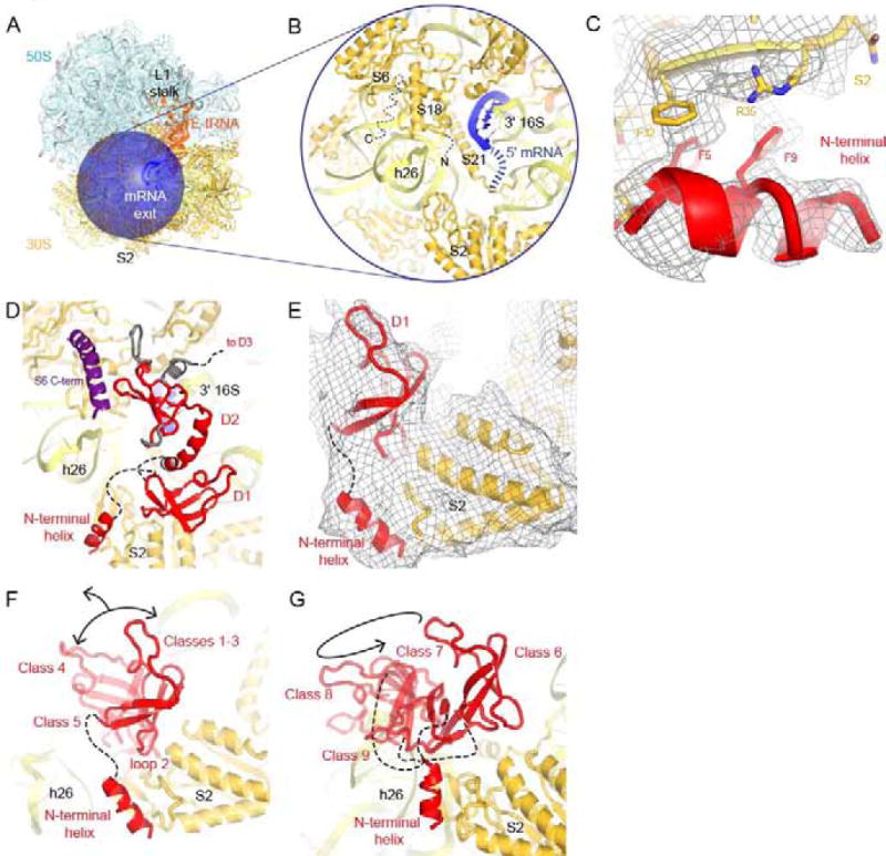
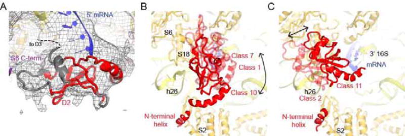
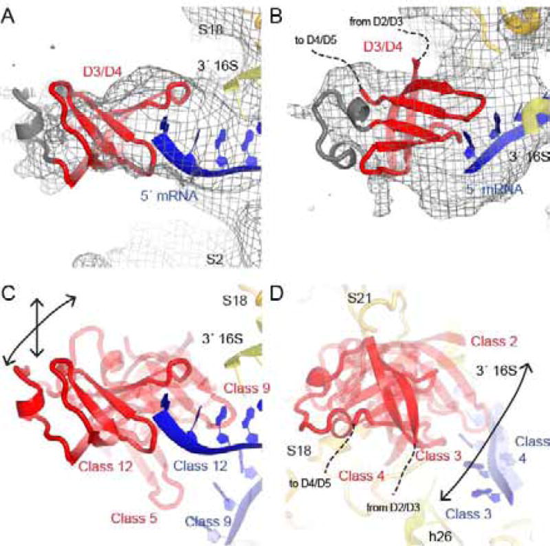
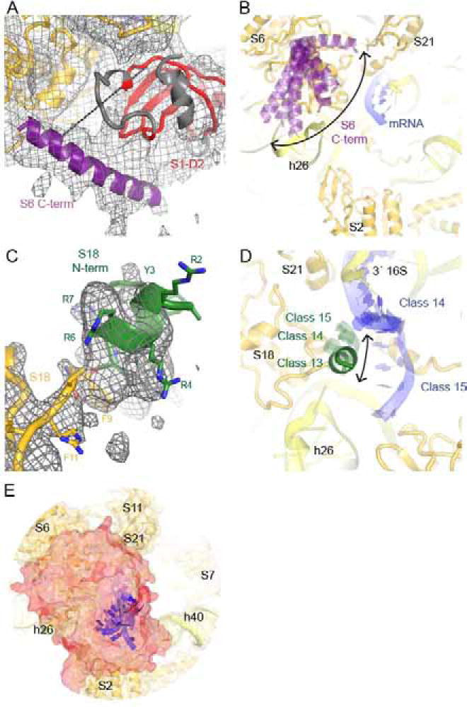
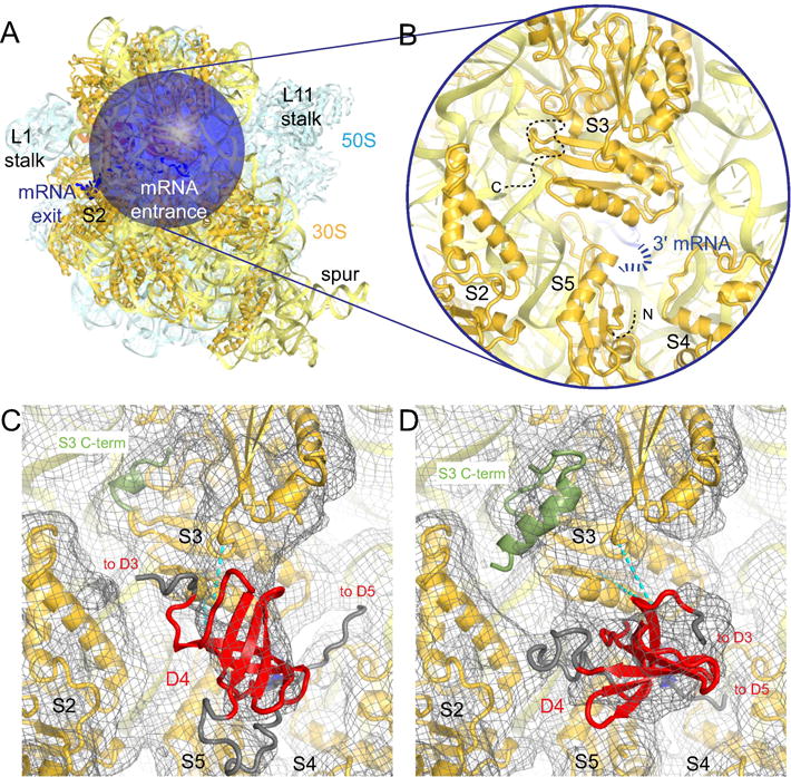
Bacterial ribosomal protein S1 is the largest and highly flexible protein of the 30S subunit, and one of a few core ribosomal proteins for which a complete structure is lacking. S1 is thought to participate in transcription and translation. Best understood is the role of S1 in facilitating translation of mRNAs with structured 5' UTRs. Here, we present cryo-EM analyses of the 70S ribosome that reveal multiple conformations of S1. Based on comparison of several 3D maximum likelihood classification approaches in Frealign, we propose a streamlined strategy for visualizing a highly dynamic component of a large macromolecular assembly that itself exhibits high compositional and conformational heterogeneity. The resulting maps show how S1 docks at the ribosomal protein S2 near the mRNA exit channel. The globular OB-fold domains sample a wide area around the mRNA exit channel and interact with mobile tails of proteins S6 and S18. S1 also interacts with the mRNA entrance channel, where an OB-fold domain can be localized near S3 and S5. Our analyses suggest that S1 cooperates with other ribosomal proteins to form a dynamic mesh near the mRNA exit and entrance channels to modulate the binding, folding and movement of mRNA.

Keywords: 3D maximum-likelihood classification; Electron cryo-microscopy; Ribosome; Rps1.


Conflict of interest statement

Competing interests

The authors declare that there are no conflicts of interest.

Figures

Figure 1
Figure 1. Approaches to visualizing S1 on the ribosome
A) Domain structure of E. coli S1. S1 domains 1 and 2 are implicated in ribosome binding while S1 domains 3–6 are required for interaction with mRNA. B) S1 adopts a wide variety of conformations on the ribosome. The pre-classification cryo-EM map is shown low-pass filtered to 10 Å and at low contour to reveal low-resolution density for unmodeled proteins L9, L1, L7/L12 and S1 (gray). Modeled density is shown in the following colors: 50S subunit (blue) and 30S subunit (gold), E- and P- site tRNA (orange). C) Global classification was used to separate particles into 4, 8, and 32 classes and revealed differences throughout the ribosome, including positions of the L1 stalk, L7/L12, L11 stalk, tRNA occupancy, intersubunit rotation (pink map), and 30S domain closure. 8 maps out of the 32-class classification are shown to highlight differences visible throughout the ribosome and the S1 binding site at S2. D) The knowledge-based approach using classification with a structure-based mask. The mask (transparent blue) was created from models of proteins that crosslink to S1 (see Methods). Separation into 8 or 48 classes revealed differences in tRNA occupancy, 30S subunit conformation (intersubunit rotation, pink map, and 30S domain closure) and density attributable to S1 near S2. 8 maps out of the 48-class classification are shown to highlight differences visible throughout the ribosome and the S1-binding site at S2. The brown class had S1 most enriched. E) The detailed classification using spherical “focus” masks (blue). A 50-Å mask was located on the 30S subunit. 8 maps out of the 16 class classifications are shown to highlight differences within the area of interest. The class lacking S1 is purple. F) The dual-mask approach using classification with spherical “focus” mask (blue) combined with a negative mask obscuring modeled regions of the ribosome (gray). The focus mask was centered at the mRNA exit channel. 8 maps out of the 16 class classifications are shown to highlight differences within the area of interest. For C–F, maps are shown filtered to 6 Å and shown at the same absolute level 0.01.
Figure 2
Figure 2. The N-terminal domain of S1 binds to S2
A) mRNA exit channel of the ribosome is shown with the focus mask used in the dual-mask approach. Colors are as in Fig. 1B. B) Close-up view of the main binding site of S1. 16S rRNA (light yellow), 30S subunit proteins (dark yellow) and 5′ end of mRNA (blue) are shown. C) Close-up view of the interaction of the N-terminal helix of S1 with S2. The cryo-EM map (Class 10) is shown at 2.5 σ after applying a B-factor of −50 Å2. D) Position of D1 in Class 1, which matches the crystal structure of the N-terminus of S1 bound to S2 (PDB:4TOI; (Byrgazov et al., 2015)). Classes 2 and 3 were also compatible with this placement for D1. E) Density for the N-terminal helix and D1 bound to S2 in Class 1. The cryo-EM map, low-pass filtered to 6 Å, is shown at 1 σ. F) Loop 2 of D1 is a second anchor point for S1 on the ribosome. In Classes 4 and 5 (both transparent), D1 was bound at S2 via loop 2, but D1 positions differ from that in Class 1 (solid red). G) D1 is detached from its main binding site at S2, while the N-terminal helix retains its position, in Classes 6–9. Classes 10 through 15 are not shown because D1 could not be placed. In panels F and G, models of D2 and D3, mRNA nucleotides 1–4 and tails of S06 and S18 were omitted for clarity.
Figure 3
Figure 3. S1 domain 2 binds in the vicinity of h26 of 16S rRNA
A) Density for D2 in the vicinity of h26 of the 16S rRNA and at the 5′ end of mRNA in Class 10. The cryo-EM map, low-pass filtered to 6 Å, is shown at 1 σ. B) D2 dynamics. In Classes 1 and 7 (transparent red), D2 is shifted compared to Class 10 (solid red). The first four nucleotides of mRNA are only ordered in Class 10 and are omitted for clarity along with D1, D3, and the C-terminal tail of S06. C) In Class 11 (solid red) and 2 (transparent red), D2 interacts with h26 by tilting relative to Class 10 (not shown). While the first four nucleotides of mRNA are ordered in Class 11, they do not contact D2 and are omitted for clarity along with D3 and the C-terminal tail of S6.
Figure 4
Figure 4. C-terminal domains of S1 are located near mRNA and the 3
end of 16S rRNA. A) An S1 domain (D3 or D4) interacts with the single-stranded 5′ nucleotides of mRNA in Class 12. The cryo-EM map, low-pass filtered to 6 Å, is shown at 1 σ. B) An alternate view of D3/D4 in Class 12. C) Like in Class 12 (solid red), D3/D4 binds near the 5′ end of mRNA in Class 9 and Class 5. The mRNA is ordered in Class 9, and not in Class 5. D) D3/D4 is located near the 3′ end of the 16S rRNA, S18 and S21 in Classes 3 (solid red), 2 and 4 (transparent red).
Figure 5
Figure 5. mRNA and S1 interact with the mobile C-terminal tail of S6 and N-terminal tail of S18
A) The C-terminal tail of S6 interacts with D2 in Class 1, consistent with crosslinking ((Lauber et al., 2012); shown by cyan dashed line). The cryo-EM map, low-pass filtered to 6 Å, is shown at 1 σ. B) Low-resolution ordered density for the C-terminal tail of S6 appears in nine Classes: 1, 3, 5, 6, 7, 8, 10, 12 and 15. C) Density for the N-terminal helix of S18 interacting with h26. The cryo-EM map (Class 13) is shown at 5 σ after applying a B-factor of −50 Å2. D) Ordered density for the N-terminal tail of S18 is also present in Classes 14 and 15, albeit at lower resolution. The tail extends towards mRNA in Classes 14 and 15 or helix 26 in Class 13. E) Superposition of S1, S6, S18 models from Classes 1 through 15 shows solid surface (red), which covers the mRNA exit channel and single-stranded mRNA positions (nucleotides 1–4 are shown). The view is as in Fig. 2A–B.
Figure 6
Figure 6. Density for S1 domains at the mRNA entrance channel
A) mRNA entrance channel of the ribosome is shown with the focus mask used in the dual-mask approach. Colors are as in Fig. 1B. B) Close-up view of the mRNA entrance channel. 16S rRNA (light yellow), 30S subunit proteins (dark yellow) and 3′ end of mRNA (blue) are shown. The flexible N- and C-terminal tails of S5 and S3, respectively, are indicated by dashed black lines. C) Density to fit an S1 domain is present at the mRNA entrance in the vicinity of the 3′ end of mRNA in Class A. The cryo-EM map, low-pass filtered to 6 Å, is shown at 0.5 σ. Cyan dashed lines indicate crosslinks observed between S3 and D4 by Lauber et. al. (Lauber et al., 2012). D) In Class B, an alternative fit of an S1 domain is possible (relative to that shown in panel C). The cryo-EM map and crosslinks are shown as in C.

Similar articles

Cited by

References

    1. Abeyrathne PD, Koh CS, Grant T, Grigorieff N, Korostelev AA. Ensemble cryo-EM uncovers inchworm-like translocation of a viral IRES through the ribosome. eLife. 2016;5:e14874. - PMC - PubMed
    1. Adams PD, Afonine PV, Bunkóczi G, Chen VB, Echols N, Headd JJ, Hung LW, Jain S, Kapral GJ, Grosse Kunstleve RW, et al. The Phenix software for automated determination of macromolecular structures. Methods (San Diego, Calif) 2011;55:94–106. - PMC - PubMed
    1. Agirrezabala X, Liao HY, Schreiner E, Fu J, Ortiz-Meoz RF, Schulten K, Green R, Frank J. Structural Characterization of mRNA-tRNA Translocation Intermediates. PNAS. 2012;109:6094–6099. - PMC - PubMed
    1. Agirrezabala X, Schreiner E, Trabuco LG, Lei J, Ortiz-Meoz RF, Schulten K, Green R, Frank J. Structural insights into cognate versus near-cognate discrimination during decoding: Unique ribosome binding of near-cognate species. The EMBO Journal. 2011;30:1497–1507. - PMC - PubMed
    1. Ahmed T, Shi J, Bhushan S. Unique localization of the plastid-specific ribosomal proteins in the chloroplast ribosome small subunit provides mechanistic insights into the chloroplastic translation. Nucleic Acids Research 2017 - PMC - PubMed

Publication types

MeSH terms

LinkOut - more resources