. 2018 Apr;154(5):1480-1493.
doi: 10.1053/j.gastro.2017.12.004. Epub 2017 Dec 14.

JTC801 Induces pH-dependent Death Specifically in Cancer Cells and Slows Growth of Tumors in Mice

Affiliations

JTC801 Induces pH-dependent Death Specifically in Cancer Cells and Slows Growth of Tumors in Mice

Xinxin Song et al. Gastroenterology. 2018 Apr.

Abstract

Background & aims: Maintenance of acid-base homeostasis is required for normal physiology, metabolism, and development. It is not clear how cell death is activated in response to changes in pH. We performed a screen to identify agents that induce cell death in a pH-dependent manner (we call this alkaliptosis) in pancreatic ductal adenocarcinoma cancer (PDAC) cells and tested their effects in mice.

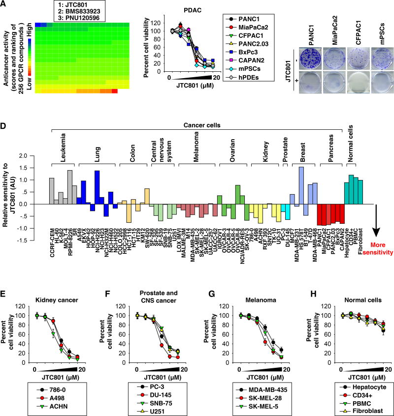
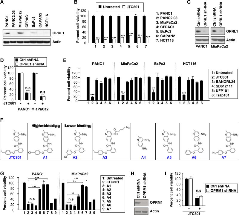
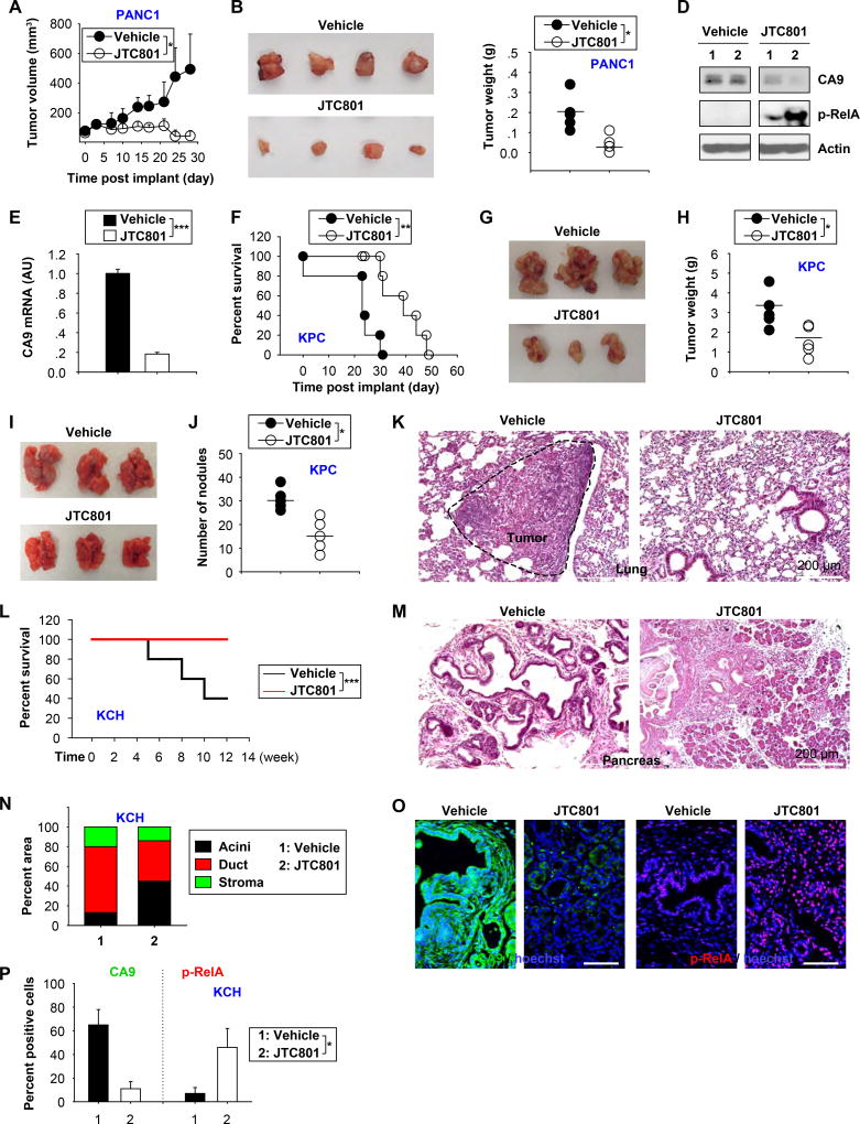
Methods: We screened a library of 254 compounds that interact with G-protein-coupled receptors (GPCRs) to identify those with cytotoxic activity against a human PDAC cell line (PANC1). We evaluated the ability of JTC801, which binds the opiod receptor and has analgesic effects, to stimulate cell death in human PDAC cell lines (PANC1, MiaPaCa2, CFPAC1, PANC2.03, BxPc3, and CAPAN2), mouse pancreatic cancer-associated stellate cell lines, primary human pancreatic ductal epithelial cells, and 60 cancer cell lines (the NCI-60 panel). Genes encoding proteins in cell death and GPCR signaling pathways, as well as those that regulate nuclear factor-κB (NF-κB) activity, were knocked out, knocked down, or expressed from transgenes in cancer cell lines. JTC801 was administered by gavage to mice with xenograft tumors, C57BL/6 mice with orthographic pancreatic tumors grown from Pdx1-Cre;KRasG12D/+;Tp53R172H/+ (KPC) cells, mice with metastases following tail-vein injection of KPC cells, and Pdx-1-Cre;KrasG12D/+ mice crossed with Hmgb1flox/flox mice (KCH mice). Pancreata were collected from mice and analyzed for tumor growth and by histology and immunohistochemistry. We compared gene and protein expression levels between human pancreatic cancer tissues and patient survival times using online R2 genomic or immunohistochemistry analyses.

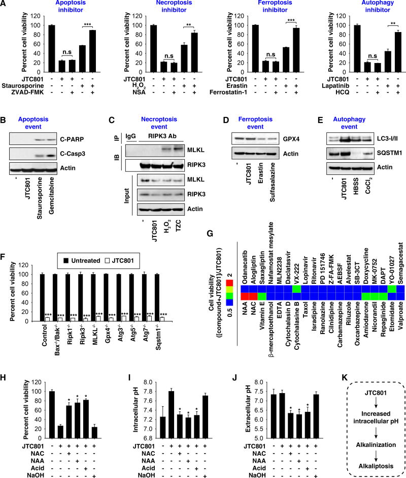
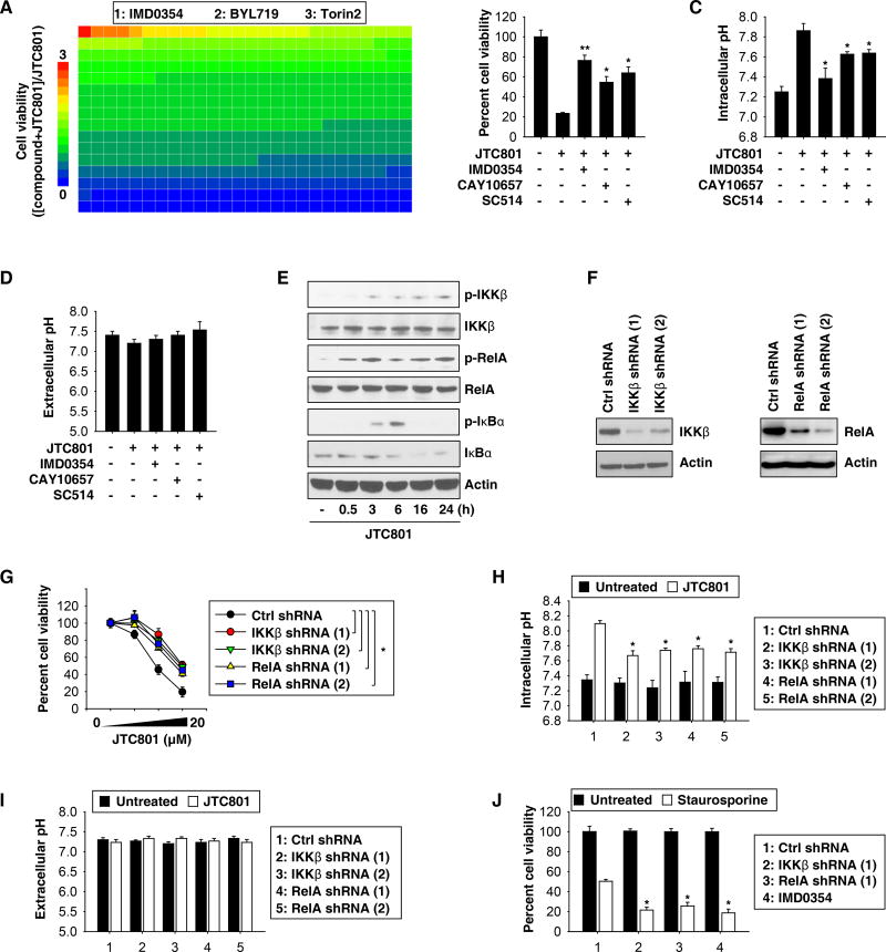
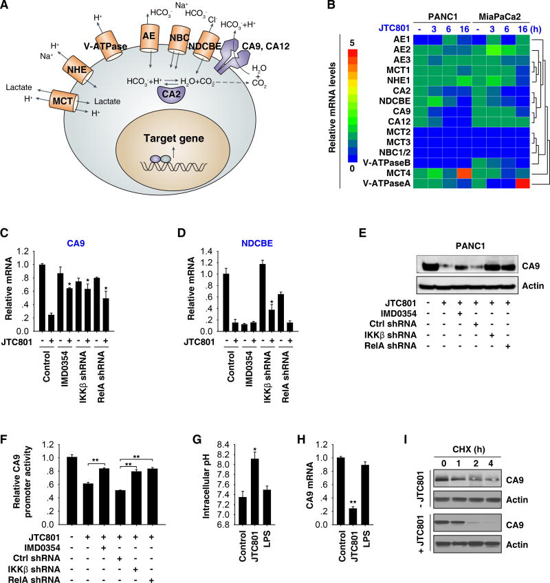
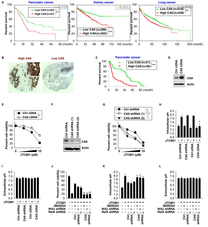
Results: Exposure of human PDAC cell lines (PANC1 and MiaPaCa2) to JTC801 did not induce molecular markers of apoptosis (cleavage of caspase 3 or poly [ADP ribose] polymerase [PARP]), necroptosis (interaction between receptor-interacting serine-threonine kinase 3 [RIPK3] and mixed lineage kinase domain like pseudokinase [MLKL]), or ferroptosis (degradation of glutathione peroxidase 4 [GPX4]). Inhibitors of apoptosis (Z-VAD-FMK), necroptosis (necrosulfonamide), ferroptosis (ferrostatin-1), or autophagy (hydroxychloroquine) did not prevent JTC801-induced death of PANC1 or MiaPaCa2 cells. The cytotoxic effects of JTC801 in immortalized fibroblast cell lines was not affected by disruption of genes that promote apoptosis (Bax-/-/Bak-/- cells), necroptosis (Ripk1-/-, Ripk3-/-, or Mlkl-/- cells), ferroptosis (Gpx4-/- cells), or autophagy (Atg3-/-, Atg5-/-, Atg7-/-, or Sqstm1-/- cells). We found JTC801 to induce a pH-dependent form cell death (alkaliptosis) in cancer cells but not normal cells (hepatocytes, bone marrow CD34+ progenitor cells, peripheral blood mononuclear cells, or dermal fibroblasts) or healthy tissues of C57BL/6 mice. JTC801 induced alkaliptosis in cancer cells by activating NF-κB, which repressed expression of the carbonic anhydrase 9 gene (CA9), whose product regulates pH balance in cells. In analyses of Cancer Genome Atlas data and tissue microarrays, we associated increased tumor level of CA9 mRNA or protein with shorter survival times of patients with pancreatic, kidney, or lung cancers. Knockdown of CA9 reduced the protective effects of NF-κB inhibition on JTC801-induced cell death and intracellular alkalinization in PANC1 and MiaPaCa2 cell lines. Oral administration of JTC801 inhibited growth of xenograft tumors (from PANC1, MiaPaCa2, SK-MEL-28, PC-3, 786-0, SF-295, HCT116, OV-CAR3, and HuH7 cells), orthotropic tumors (from KPC cells), lung metastases (from KPC cells) of mice, and slowed growth of tumors in KCH mice.

Conclusions: In a screen of agents that interact with GPCR pathways, we found JTC801 to induce pH-dependent cell death (alkaliptosis) specifically in cancer cells such as PDAC cells, by reducing expression of CA9. Levels of CA9 are increased in human cancer tissues. JTC801 might be developed for treatment of pancreatic cancer.

Keywords: Drug Development; Pancreas; Targeted Therapy; Tumor Microenvironment.


Conflict of interest statement

Disclosure of Potential Conflicts of Interest: No potential conflicts of interest were disclosed.

Figures

Fig. 1
Fig. 1. Anticancer activity of JTC801 in vitro
(A) PANC1 cells were treated with a GPCR compound (10 µM) for 24 hours and then cell viability was assayed. The ranking of the relative anticancer activity of 254 GPCR compounds is shown by the heat map; each block represents one GPCR compound. The top three anti-cancer GPCR compounds are listed. (B) Indicated human PDAC cell lines, mPSCs, and hPDEs were treated with JTC801 (1.25–20 µM) for 24 hours. Cell viability was assayed (n=3). (C) Clonogenic cell survival assay determined the reproductive ability of a cell in response to JTC801 (10 µM). (D) Sensitivity profile of 66 cancer cell lines and four normal cell types against JTC801. The cells were grouped based on their tissue origins. (E–H) Indicated cancer or normal cells were treated with JTC801 (1.25–20 µM) for 24 hours. Cell viability was assayed (n=3).
Fig. 2
Fig. 2. JTC801 induces alkaliptosis
(A) PANC1 cells were treated with JTC801 (10 µM), staurosporine (0.5 µM), erastin (20 µM), H2O2 (500 µM), or lapatinib (50 µM) in the absence or presence of ZVAD-FMK (20 µM), ferrostatin-1 (500 nM), necrosulfonamide (“NSA”, 1 µM), and hydroxychloroquine (“HCQ”, 50 µM) for 24 hours. Cell viability was assayed (n=3, **p < 0.01, ***p < 0.001, n.s.=not significant). (B) PANC1 cells were treated with JTC801 (10 µM), staurosporine (1 µM), or gemcitabine (5 mM) for 24 hours and the levels of cleaved-PARP (“C-PARP”), cleaved-caspase 3 (“C-Casp3”), and actin were assayed using western blot. (C) PANC1 cells were treated with JTC801 (10 µM), H2O2 (500 µM), or TZC (TNF [50 nM]/ZVAD-FMK [20 µM]/cycloheximide [10 µg/ml]) for 24 hours. Cell lysates were immunoprecipitated with anti-RIPK3 antibody or control IgG, and then the levels of MLKL and RIPK3 were assayed using western blot. (D) PANC1 cells were treated with JTC801 (10 µM), erastin (20 µM), or salazosulfapyridine (1 mM) for 24 hours and then the levels of GPX4 and actin were assayed using western blot. (E) PANC1 cells were treated with JTC801 (10 µM, 24 hours), CoCl2 (400 µM, 24 hours), or Hank's buffered salt solution (HBSS, 6 hours), and the levels of LC3-I/-II, SQSTM1, and actin were assayed using western blot. (F) Indicated gene-deficient cells were treated with JTC801 (10 µM) for 24 hours, and then cell viability was assayed (n=3, ***p < 0.001 versus untreated group). (G) PANC1 cells were treated with JTC801 (10 µM) in the absence or presence of the indicated compounds (10 µM) for 24 hours, and then cell viability was assayed. The ratio in the heat map was defined as cell viability ([Compound+JTC801]/JTC801). (H–J) PANC1 cells were treated with JTC801 (10 µM) in the absence or presence of N-acetylcysteine (“NAC”, 100 mM), N-acetyl alanine acid (“NAA”, 100 mM), acidic medium (pH=6, adjusted by HCl), and NAC (100 mM, pH=7, adjusted by NaOH) for 24 hours. Cell viability, intracellular pH, and extracellular pH were assayed (n=3, *p < 0.05 versus JTC801 group). (K) Schematic depicting JTC801-induced alkaliptosis.
Fig. 3
Fig. 3. OPRL1 is not required for JTC801-induced alkaliptosis
(A) Western blot analysis of OPRL1 expression in cancer cells. (B) Cancer cells were treated with JTC801 (10 µM) for 24 hours and then cell viability was assayed (n=3, ***p < 0.001 versus untreated group). (C) Western blot analysis of OPRL1 expression in OPRL1-knockdown PANC1 and MiaPaCa2 cells. (D) Knockdown of OPRL1 did not affect JTC801 (10 µM, 24 hours)-induced cell death in PANC1 and MiaPaCa2 cells (n=3, n.s=not significant). (E) Cancer cells were treated with JTC801 or indicated OPRL1 antagonists at 10 µM for 24 hours. Cell viability was assayed (n=3, **p < 0.01, ***p < 0.001 versus untreated group). (F) Structure of JTC801 and its analogues. (G) PANC1 and MiaPaCa2 cells were treated with JTC801 and its analogues at 10 µM for 24 hours. Cell viability was assayed (n=3, **p < 0.01, ***p < 0.001, n.s=not significant). (H) Western blot analysis of OPRM1 expression in OPRL1-knockdown PANC1 cells. (I) Knockdown of OPRM1 did not affect JTC801 (10 µM, 24 hours)-induced cell death in PANC1 cells (n=3, n.s=not significant).
Fig. 4
Fig. 4. Activation of NF-κB contributes to alkaliptosis
(A) PANC1 cells were treated with JTC801 (10 µM) in the absence or presence of an inhibitor (10 µM) for 24 hours and then cell viability was assayed. The relative effects of 416 inhibitors on anticancer activity of JTC801 are shown by the heat map. The top three inhibitors of the anticancer activity of JTC801 are listed. (B–D) PANC1 cells were treated with JTC801 (10 µM) in the absence or presence of indicated IKKβ inhibitors (10 µM) for 24 hours. Cell viability, intracellular pH, and extracellular pH were measured (n=3, *p < 0.05, **p < 0.01 versus JTC801 group). (E) Western blot analysis of expressions of indicated proteins in PANC1 cells in response to JTC801 (10 µM) for 0.5–24 hours. (F) Western blot analysis of IKKβ and RelA expression in indicated gene-knockdown PANC1 cells. (G) Knockdown of IKKβ and RelA inhibited JTC801 (1.25–20 µM)-induced cell death at 24 hours in PANC1 cells (n=3, *p < 0.05). (H–I) Indicated gene-knockdown PANC1 cells were treated with JTC801 (10 µM) for 24 hours. Intracellular and extracellular pH was assayed (n=3, *p < 0.05 versus untreated group). (J) Indicated gene-knockdown PANC1 cells were treated with staurosporine (1 µM) with or without IMD0354 (10 µM) for 24 hours. Cell viability was measured (n=3, *p < 0.05 versus control shRNA group).
Fig. 5
Fig. 5. CA9 is a negative target of NF-κB in alkaliptosis
(A) Schematic depicting proteins involved in pH regulation within a tumor cell. (B) PANC1 and MiaPaCa2 cells were treated with JTC801 (10 µM) for three to 16 hours and then the indicated genes were assayed using Q-PCR. The relative mRNA levels of indicated genes are shown by the heat map. (C–D) Q-PCR analysis of mRNA levels of CA9 and NDCBE in indicated PANC1 cells in response to JTC801 (10 µM) in the absence or presence IMD0354 (10 µM) for 24 hours (n=3, *p < 0.05 versus control JTC801 group). (E–F) In parallel, the protein expression and promoter luciferase activity of CA9 were measured (n=3, **p < 0.01). (G–H) PANC1 were treated with JTC801 (10 µM) or LPS (500 ng/ml) for 16 hours and then intracellular pH and CA9 mRNA were assayed (n=3, *p < 0.05, **p < 0.01 versus control group). (I) PANC1 cells were treated with JTC801 (10 µM) and cycloheximide (CHX, 20 µg/ml) for indicated time points. Cell lysates were subjected to western blot analysis with anti-CA9 and anti-actin antibodies.
Fig. 6
Fig. 6. CA9 downregulation contributes to alkaliptosis
(A) Survival analysis of patients with low or high CA9 mRNA expression levels in pancreatic, kidney, and lung cancer from the SurvExpress online gene expression database (http://bioinformatica.mty.itesm.mx:8080/Biomatec/SurvivaX.jsp; **p < 0.01, ***p < 0.001). (B, C) Higher level of CA9 expression was associated with shorter survival times of patients with pancreatic cancer. Representative tissue microarray spots stained for higher or lower CA9 expression by immunohistochemistry are shown in panel B (***p < 0.001). (D) Western blot analysis of CA9 expression in CA9-overexpression PANC1 cells. (E) Overexpression of CA9 inhibited JTC801-induced cell death at 24 hours in PANC1 cells (n=3, *p < 0.05). (F) Western blot analysis of CA9 expression in CA9-knockdown PANC1 cells. (G) Knockdown of CA9 increased JTC801-induced cell death at 24 hours in PANC1 cells (n=3, *p < 0.05 versus control shRNA group). (H, I) Intracellular and extracellular pH was assayed in indicated PANC1 cells in response to JTC801 (10 µM) for 24 hours (n=3, *p < 0.05 versus control group). (J–L) Knockdown of CA9 restored JTC801-induced cell death in NF-κB-inhibition (IMD0354 [10 µM]) or - knockdown (IKKβ shRNA or RelA shRNA) PANC1 cells. In parallel, intracellular and extracellular pH was assayed (n=3, *p < 0.05, **p < 0.01 versus control shRNA group).
Fig. 7
Fig. 7. Anticancer activity of JTC801 in vivo
(A) Oral injection of JTC801 (20 mg/kg, once every day, at day seven for two weeks) inhibited tumor growth in a xenograft model of PANC1 tumor models (n=8 mice/group, * p < 0.05). (B) Representative photomicrographs of isolated tumors at day 28. (C–D) In parallel, tumor weight (C), indicated protein expression (D), and CA9 mRNA (E) were assayed at day 28 (n=5 mice/group, * p < 0.05, *** p < 0.001). (F) Oral injection of JTC801 (20 mg/kg, once every day, at day seven for three weeks) prolonged C57BL/6 mouse survival of orthotopic KPC tumor models (n=10 mice/group, ** p < 0.01). (G) Representative photomicrographs of isolated tumors at day 26. (H) In parallel, tumor weights were assayed at day 26 (n=5 mice/group, * p < 0.05). (I) Oral injection of JTC801 (20 mg/kg, once every day, at day seven for two weeks) limited the formation of lung metastasis in C57BL/6 mice based on tail vein injection of KPC cells. (J–K) The number of tumor nodules (J) and histology (K) in lung were assayed at day 28 (n=5 mice/group, * p < 0.05). (L) Oral injection of JTC801 (20 mg/kg, twice per week, started at four weeks of age for four weeks) prolonged survival in KCH mice at 12 weeks of age (n=10 mice/group, *** p < 0.001). (M–P) In parallel, pancreatic histology (M, N) and relative expression of CA9 and p-RelA in the pancreas (O, P) were assayed (n=5 mice/group, * p < 0.05).

References

    1. Galluzzi L, Bravo-San Pedro JM, Vitale I, et al. Essential versus accessory aspects of cell death: recommendations of the NCCD 2015. Cell Death Differ. 2014 - PMC - PubMed
    1. Hanahan D, Weinberg RA. Hallmarks of cancer: the next generation. Cell. 2011;144:646–74. - PubMed
    1. O'Hayre M, Vazquez-Prado J, Kufareva I, et al. The emerging mutational landscape of G proteins and G-protein-coupled receptors in cancer. Nat Rev Cancer. 2013;13:412–24. - PMC - PubMed
    1. Pikarsky E, Porat RM, Stein I, et al. NF-kappaB functions as a tumour promoter in inflammation-associated cancer. Nature. 2004;431:461–6. - PubMed
    1. Xie Y, Zhu S, Zhong M, et al. Inhibition of Aurora Kinase A Induces Necroptosis in Pancreatic Carcinoma. Gastroenterology. 2017;153:1429–1443. e5. - PMC - PubMed

Publication types

MeSH terms