

Metaplastic Cells in the Stomach Arise, Independently of Stem Cells, via Dedifferentiation or Transdifferentiation of Chief Cells
- PMID: 29248442
- PMCID: PMC5847468
- DOI: 10.1053/j.gastro.2017.11.278
Metaplastic Cells in the Stomach Arise, Independently of Stem Cells, via Dedifferentiation or Transdifferentiation of Chief Cells
Abstract
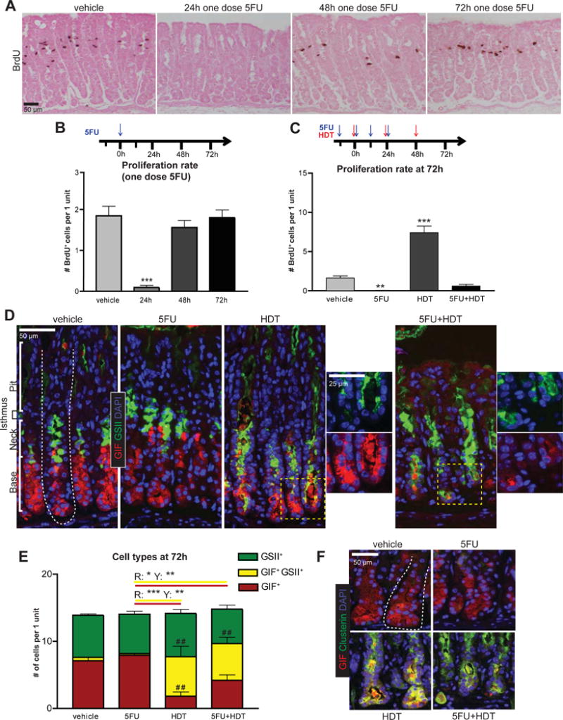
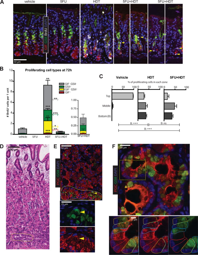
Spasmolytic polypeptide-expressing metaplasia (SPEM) develops in patients with chronic atrophic gastritis due to infection with Helicobacter pylori; it might be a precursor to intestinal metaplasia and gastric adenocarcinoma. Lineage tracing experiments of the gastric corpus in mice have not established whether SPEM derives from proliferating stem cells or differentiated, post-mitotic zymogenic chief cells in the gland base. We investigated whether differentiated cells can give rise to SPEM using a nongenetic approach in mice. Mice were given intraperitoneal injections of 5-fluorouracil, which blocked gastric cell proliferation, plus tamoxifen to induce SPEM. Based on analyses of molecular and histologic markers, we found SPEM developed even in the absence of cell proliferation. SPEM therefore did not arise from stem cells. In histologic analyses of gastric resection specimens from 10 patients with adenocarcinoma, we found normal zymogenic chief cells that were transitioning into SPEM cells only in gland bases, rather than the proliferative stem cell zone. Our findings indicate that SPEM can arise by direct reprogramming of existing cells-mainly of chief cells.
Keywords: Cancer; Development; Differentiation; Exocrine Cell.
Copyright © 2018 AGA Institute. Published by Elsevier Inc. All rights reserved.
Conflict of interest statement
The authors disclose no conflicts.
Figures
Comment in
-
Cellular Plasticity in the Stomach: Insights Into the Cellular Origin of Gastric Metaplasia.Gastroenterology. 2018 Mar;154(4):801-803. doi: 10.1053/j.gastro.2018.02.001. Epub 2018 Feb 6. Gastroenterology. 2018. PMID: 29425924 Free PMC article. No abstract available.
References
-
- Matsuo J, et al. Gastroenterology. 2017;152:218–231.e14. - PubMed
Publication types
MeSH terms
Substances
Grants and funding
LinkOut - more resources
Full Text Sources
Other Literature Sources
Medical
