. 2018 Apr:67:90-106.
doi: 10.1016/j.matbio.2017.12.003. Epub 2017 Dec 14.

Extracellular matrix directs phenotypic heterogeneity of activated fibroblasts

Affiliations

Extracellular matrix directs phenotypic heterogeneity of activated fibroblasts

Diana Avery et al. Matrix Biol. 2018 Apr.

Abstract

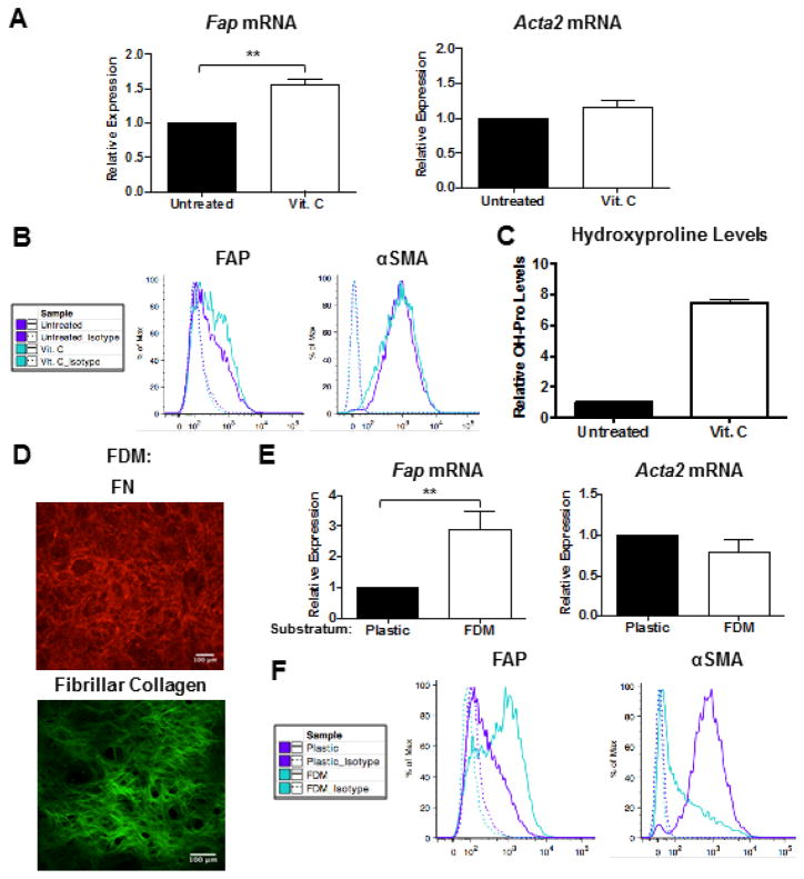
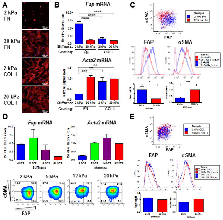
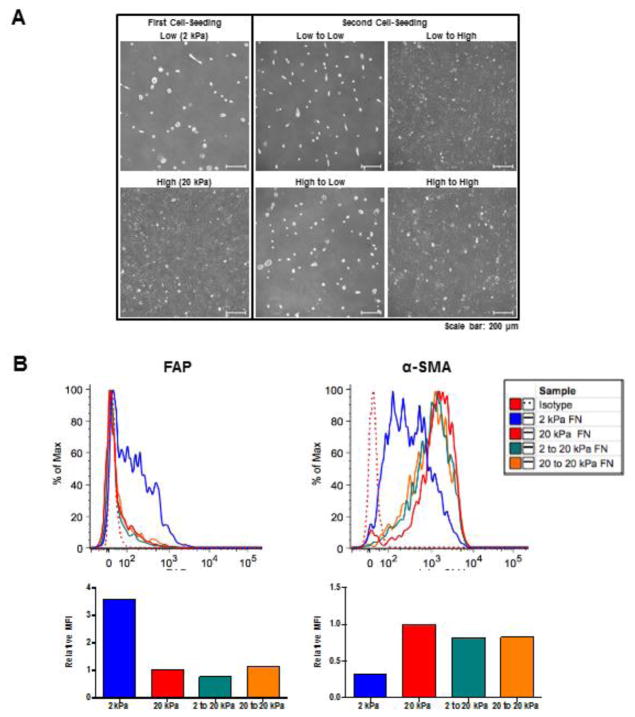
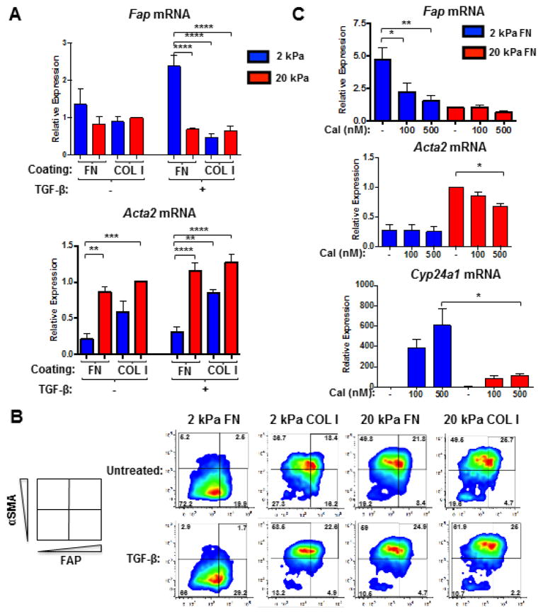
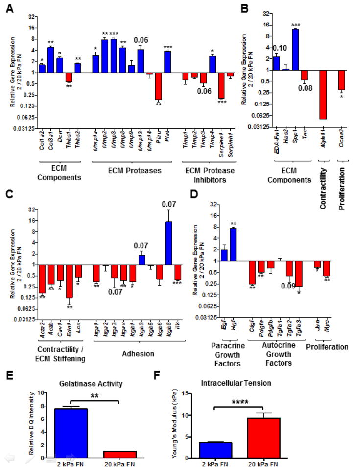
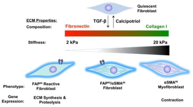
Activated fibroblasts are key players in the injury response, tumorigenesis, fibrosis, and inflammation. Dichotomous outcomes in response to varied stroma-targeted therapies in cancer emphasize the need to disentangle the roles of heterogeneous fibroblast subsets in physiological and pathophysiological settings. In wound healing, fibrosis, and myriad tumor types, fibroblast activation protein (FAP) and alpha-smooth muscle actin (αSMA) identify distinct, yet overlapping, activated fibroblast subsets. Prior studies established that FAPHi reactive fibroblasts and αSMAHi myofibroblasts can exert opposing influences in tumorigenesis. However, the factors that drive this phenotypic heterogeneity and the unique functional roles of these subsets have not been defined. We demonstrate that a convergence of ECM composition, elasticity, and transforming growth factor beta (TGF-β) signaling governs activated fibroblast phenotypic heterogeneity. Furthermore, FAPHi reactive fibroblasts and αSMAHi myofibroblasts exhibited distinct gene expression signatures and functionality in vitro, illuminating potentially unique roles of activated fibroblast subsets in tissue remodeling. These insights into activated fibroblast heterogeneity will inform the rational design of stroma-targeted therapies for cancer and fibrosis.

Keywords: Activated fibroblasts; Alpha-smooth muscle actin; Fibroblast activation protein; Fibroblast heterogeneity.


Figures

Figure 1
Figure 1. Substratum directs activated fibroblast phenotypic heterogeneity
QRT-PCR (A) and representative flow cytometric analysis (B) of FAP and αSMA expression in fibroblasts cultured in 10% serum on tissue culture-treated plastic in the presence or absence of 75 μg/ml ascorbic acid (Vit. C) for 4 days. Data were compiled from 4 independent experiments and bar graphs depict the mean +/− SEM. (C) Collagen levels (as measured via hydroxyproline content) in FDMs deposited by fibroblasts in the presence or absence of 75 μg/ml Vit. C. Data were compiled from 2 independent experiments and bar graphs depict the mean +/− SEM. (D) Representative IF staining of FN and two-photon second harmonic generation imaging of fibrillar collagen in lung FDMs. QRT-PCR (E) and representative flow cytometric analysis (F) of FAP and αSMA expression in fibroblasts in 10% serum on tissue culture-treated plastic or FDM for 4 days. Data were compiled from 4 independent experiments and bar graphs depict the mean +/− SEM.
Figure 2
Figure 2. ECM composition and elasticity govern activated fibroblast phenotypic heterogeneity
Representative phalloidin staining of the actin cytoskeleton (A) and Fap and Acta2 gene expression (B) in fibroblasts cultured in 10% serum on 2 versus 20 kPa FN- or COL I-coated hydrogels for 72 hours. Data were compiled from 4 independent experiments and bar graphs depict the mean +/− SEM. (C) Representative flow cytometric analysis, including quantification of relative median fluorescent intensities (MFI) for FAP and αSMA expression in fibroblasts cultured in 10% serum on 2 kPa (blue) versus 20 kPa (red) FN-coated hydrogels for 72 hours. Data were compiled from 3 independent experiments and bar graphs depict the mean +/− SEM. (D) QRT-PCR (top) and flow cytometric analysis (bottom) of FAP and αSMA expression in fibroblasts cultured in 10% serum on 2, 5, 12, and 20 kPa FN-coated hydrogels for 72 hours. Data were compiled from 2 independent experiments and bar graphs depict the mean +/− SEM. (E) Representative flow cytometric analysis, including quantification of relative MFI for FAP and αSMA expression in lung fibroblasts cultured in 10% serum on 2 kPa (blue) versus 20 kPa (red) COL I-coated hydrogels for 72 hours. Data were compiled from 3 independent experiments and bar graphs depict the mean +/− SEM.
Figure 3
Figure 3. FAPHi and αSMAHi fibroblasts exhibit morphologic and phenotypic plasticity
Fibroblasts were serially cultured (in 10% FCS DMEM) first on either 2 or 20 kPa FN-coated hydrogels for 72 hours, and then cells recovered by trypsinization from each of these groups were cultured for a second time on either 2 or 20 kPa FN-coated hydrogels for an additional 72 hours. At both rounds 1 and 2, cell monolayers were analyzed by phase contrast microscopy and cell suspensions were analyzed by flow cytometry. Representative phase contrast images (A) and flow cytometric analysis (B), including quantification of relative MFI for FAP and αSMA expression, are shown.
Figure 4
Figure 4. Fibroblast differentiation in response to TGF-β is governed by ECM composition and elasticity
QRT-PCR (A) and flow cytometric analysis (B) of FAP and αSMA expression in fibroblasts that were seeded in 1% serum on 2 versus 20 kPa COL I- or FN-coated hydrogels, allowed to adhere overnight, and subsequently treated with 2 ng/ml TGF-β for 48 (A) or 72 hours (B). Data were compiled from 4 independent experiments and bar graphs depict the mean +/− SEM. (C) QRT-PCR analysis of Fap, Acta2, and Cyp24a1 gene expression in fibroblasts that were seeded in 10% serum on FN-coated hydrogels (2 or 20 kPa), allowed to adhere overnight, and subsequently treated with calcipotriol (Cal; 100 or 500 nM) or vehicle control for 48 hours. Data were compiled from 3 independent experiments and bar graphs depict the mean +/− SEM.
Figure 5
Figure 5. Gene expression profiling and functional tests indicate that FAPHi reactive fibroblasts predominantly synthesize and proteolyze ECM, while αSMAHi myofibroblasts mediate contraction
Comparison of gene expression profiles, assessed via Qiagen Fibrosis Gene Expression Array (A, C, and D) and independent qRT-PCR analyses (B), of fibroblasts cultured in 10% serum on 2 (FAPHi fibroblasts) or 20 kPa (αSMAHi fibroblasts) FN-coated hydrogels for 72 hours. Relative gene expression (FAPHi/αSMAHi fibroblasts) of ECM proteases (A), ECM protease inhibitors (A), ECM and matricellular components (A and B), contractile mediators (B and C), integrin subunits (C), growth factors (D), and proliferative mediators (B and D). Data were compiled from 3 independent experiments and bar graphs depict the mean +/− SEM. (E) Quantification of relative DQ intensity of fibroblasts that were cultured on 2 or 20 kPa FN-coated hydrogels for 48 hours, and then incubated with DQ gelatin for an additional 24 hours. Data were compiled from 3 independent experiments and bar graphs depict the mean +/− SEM. (F) Atomic force microscopy (AFM) measurements of intracellular tension in fibroblasts cultured in 10% serum on 2 or 20 kPa FN-coated hydrogels for 72 hours. Bar graph depicts the mean +/− SEM (n=10 cells).
Figure 6
Figure 6. A convergence of ECM composition, elasticity, and TGF-β signaling governs activated fibroblast phenotypic heterogeneity
In FN-rich ECMs of low stiffness, TGF-β promotes the FAPHiαSMALow reactive fibroblast phenotype. Conversely, TGF-β promotes the FAPLowαSMAHi myofibroblast phenotype in the context of stiff and/or COL I-rich ECMs. Gene expression profiling indicates unique functionality of these divergent fibroblast subsets in tissue remodeling, with FAPHi reactive fibroblasts exhibiting an ECM synthetic and proteolytic phenotype, and αSMAHi myofibroblasts exhibiting a contractile and proliferative phenotype.

References

    1. Servais C, Erez N. From sentinel cells to inflammatory culprits: cancer-associated fibroblasts in tumour-related inflammation. J Pathol. 2013;229:198–207. doi: 10.1002/path.4103. - DOI - PubMed
    1. Tomasek JJ, Gabbiani G, Hinz B, Chaponnier C, Brown RA. Myofibroblasts and mechano-regulation of connective tissue remodelling. Nat Rev Mol Cell Biol. 2002;3:349–363. http://dx.doi.org/10.1038/nrm809. - DOI - PubMed
    1. Hinz B, Phan SH, Thannickal VJ, Prunotto M, Desmoulière A, Varga J, De Wever O, Mareel M, Gabbiani G. Recent developments in myofibroblast biology: paradigms for connective tissue remodeling. Am J Pathol. 2012;180:1340–1355. doi: 10.1016/j.ajpath.2012.02.004. - DOI - PMC - PubMed
    1. Hinz B, Phan SH, Thannickal VJ, Galli A, Bochaton-Piallat M-L, Gabbiani G. The myofibroblast: one function, multiple origins. Am J Pathol. 2007;170:1807–1816. doi: 10.2353/ajpath.2007.070112. - DOI - PMC - PubMed
    1. Serini G, Gabbiani G. Mechanisms of myofibroblast activity and phenotypic modulation. Exp Cell Res. 1999;250:273–283. doi: 10.1006/excr.1999.4543. - DOI - PubMed

Publication types