

miR-200a-5p regulates myocardial necroptosis induced by Se deficiency via targeting RNF11
- PMID: 29248830
- PMCID: PMC5975215
- DOI: 10.1016/j.redox.2017.11.025
miR-200a-5p regulates myocardial necroptosis induced by Se deficiency via targeting RNF11
Abstract
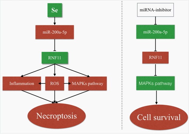
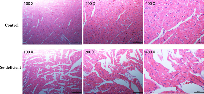
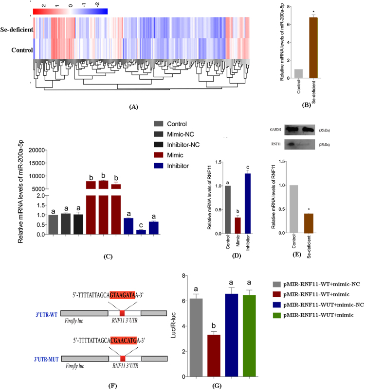
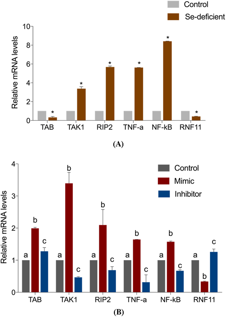
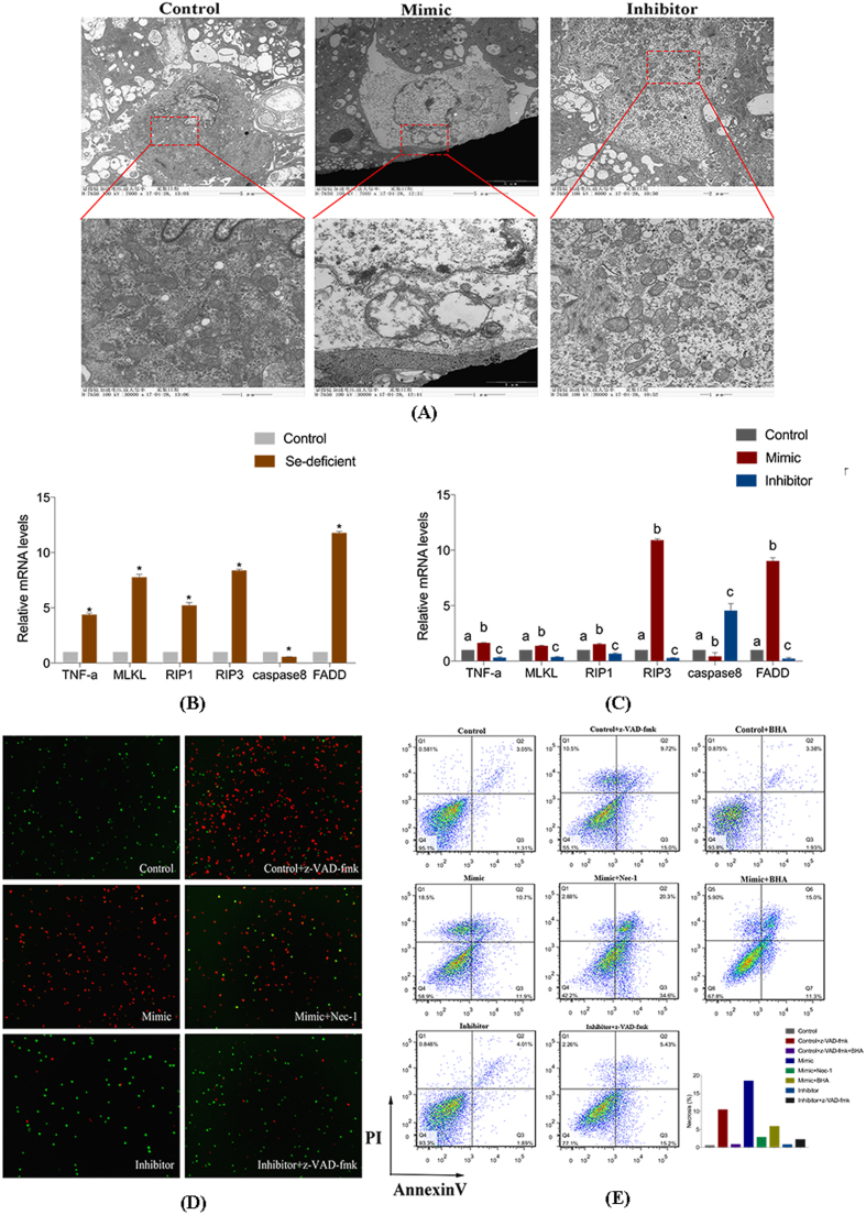
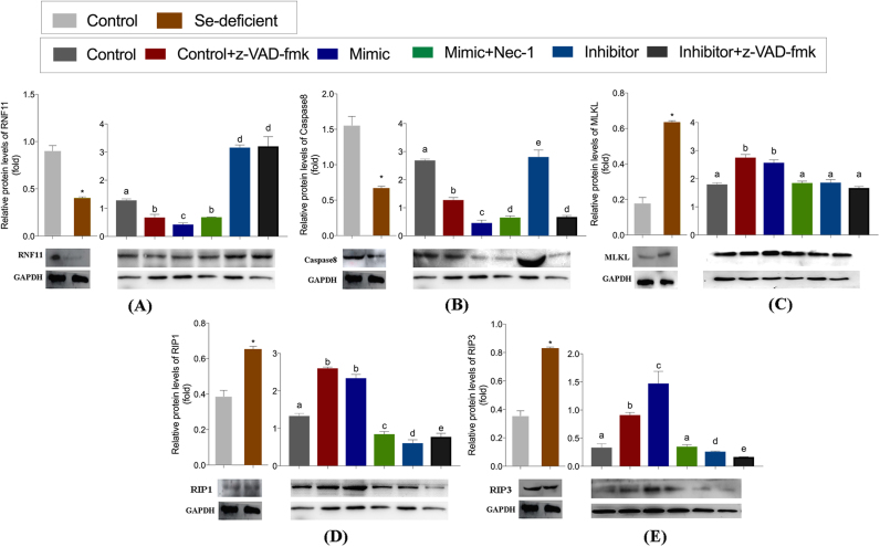
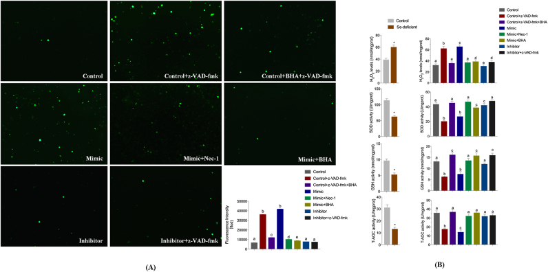
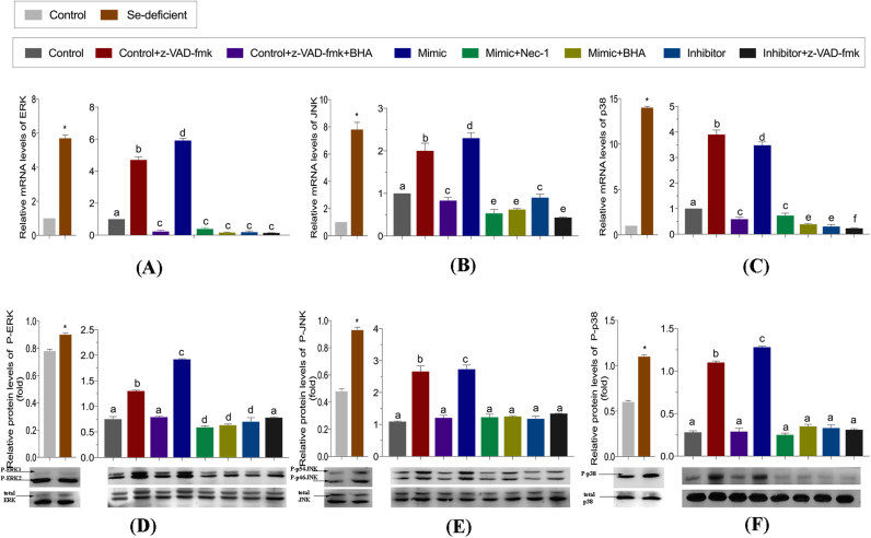
Necroptosis has been discovered as a new paradigm of cell death and may play a key role in heart disease and selenium (Se) deficiency. Hence, we detected the specific microRNA (miRNA) in response to Se-deficient heart using microRNAome analysis. For high-throughput sequencing using Se-deficient chicken cardiac tissue, we selected miR-200a-5p and its target gene ring finger protein 11 (RNF11) based on differential expression in cardiac tissue and confirmed the relationship between miR-200a-5p and RNF11 by dual luciferase reporter assay and real-time quantitative PCR (qRT-PCR) in cardiomyocytes. We further explored the function of miR-200a-5p and observed that overexpression of miR-200a-5p spark the receptor interacting serine/threonine kinase 3 (RIP3)-dependent necroptosis in vivo and in vitro. To understand whether miR-200a-5p and RNF11 are involved in the RIP3-dependent necroptosis pathway, we presumed that oxidative stress, inflammation response and the mitogen-activated protein kinase (MAPK) pathway might trigger necroptosis. Interestingly, necroptosis trigger, z-VAD-fmk, failed to induce necroptosis but enhanced cell survival against necrosis in cardiomyocytes with knockdown of miR-200a-5p. Our present study provides a new insight that the modulation of miR-200a-5p and its target gene might block necroptosis in the heart, revealing a novel myocardial necrosis regulation model in heart disease.
Keywords: Cardiomyocytes; Necroptosis; RNF11; Selenium; miR-200a-5p.
Copyright © 2018 The Authors. Published by Elsevier B.V. All rights reserved.
Figures
Similar articles
-
Mir-215-5p induces autophagy by targeting PI3K and activating ROS-mediated MAPK pathways in cardiomyocytes of chicken.J Inorg Biochem. 2019 Apr;193:60-69. doi: 10.1016/j.jinorgbio.2019.01.010. Epub 2019 Jan 18. J Inorg Biochem. 2019. PMID: 30684759
-
Bisphenol A Regulates the TNFR1 Pathway and Excessive ROS Mediated by miR-26a-5p/ADAM17 Axis to Aggravate Selenium Deficiency-Induced Necroptosis in Broiler Veins.Biol Trace Elem Res. 2024 Apr;202(4):1722-1740. doi: 10.1007/s12011-023-03756-3. Epub 2023 Jul 8. Biol Trace Elem Res. 2024. PMID: 37422542
-
Selenium deficiency exacerbates LPS-induced necroptosis by regulating miR-16-5p targeting PI3K in chicken tracheal tissue.Metallomics. 2020 Apr 1;12(4):562-571. doi: 10.1039/c9mt00302a. Epub 2020 Mar 3. Metallomics. 2020. PMID: 32125337
-
Selenium deficiency inhibits myocardial development and differentiation by targeting the mir-215-5p/CTCF axis in chicken.Metallomics. 2019 Feb 20;11(2):415-428. doi: 10.1039/c8mt00319j. Metallomics. 2019. PMID: 30565618
-
MiR-128-1-5p regulates tight junction induced by selenium deficiency via targeting cell adhesion molecule 1 in broilers vein endothelial cells.J Cell Physiol. 2018 Nov;233(11):8802-8814. doi: 10.1002/jcp.26794. Epub 2018 Jun 15. J Cell Physiol. 2018. PMID: 29904913
Cited by
-
The Role of Selenium in Atherosclerosis Development, Progression, Prevention and Treatment.Biomedicines. 2023 Jul 17;11(7):2010. doi: 10.3390/biomedicines11072010. Biomedicines. 2023. PMID: 37509649 Free PMC article. Review.
-
Selenium Deficiency Induces Apoptosis and Necroptosis Through ROS/MAPK Signal in Human Uterine Smooth Muscle Cells.Biol Trace Elem Res. 2022 Jul;200(7):3147-3158. doi: 10.1007/s12011-021-02910-z. Epub 2021 Sep 4. Biol Trace Elem Res. 2022. PMID: 34480665
-
Inhibition of gap junction composed of Cx43 prevents against acute kidney injury following liver transplantation.Cell Death Dis. 2019 Oct 10;10(10):767. doi: 10.1038/s41419-019-1998-y. Cell Death Dis. 2019. PMID: 31601792 Free PMC article.
-
Non-coding RNAs in necroptosis, pyroptosis, and ferroptosis in cardiovascular diseases.Front Cardiovasc Med. 2022 Aug 4;9:909716. doi: 10.3389/fcvm.2022.909716. eCollection 2022. Front Cardiovasc Med. 2022. PMID: 35990979 Free PMC article. Review.
-
Principal component analysis of blood microRNA datasets facilitates diagnosis of diverse diseases.PLoS One. 2020 Jun 5;15(6):e0234185. doi: 10.1371/journal.pone.0234185. eCollection 2020. PLoS One. 2020. PMID: 32502186 Free PMC article.
References
-
- Eriksson L.C. Selenium homeostasis and thioredoxin reductase induction at long term treatment with selenite in tumor preventive doses. Adv. Funct. Mater. 2006;20(3):377–385.
-
- Yusuf S.W., Rehman Q., Casscells W. Cardiomyopathy in association with selenium deficiency: a case report. J. Parenter. Enter. Nutr. 2002;26(1):63. - PubMed
-
- Li C. Selenium deficiency and endemic heart failure in China: a case study of biogeochemistry for human health. Ambio. 2013;36(1):90. - PubMed
-
- Pallarés F.J. Vitamin E and selenium concentrations in livers of pigs diagnosed with mulberry heart disease. J. Vet. Diagn. Investig. 2002;14(5):412. - PubMed
-
- Lymbury R.S., Marino M.J., Perkins A.V. Effect of dietary selenium on the progression of heart failure in the ageing spontaneously hypertensive rat. Mol. Nutr. Food Res. 2010;54(10):1436. - PubMed
Publication types
MeSH terms
Substances
LinkOut - more resources
Full Text Sources
Other Literature Sources
Miscellaneous
