. 2018 Apr;67(4):525-536.
doi: 10.1007/s00262-017-2105-9. Epub 2017 Dec 16.

Human c-SRC kinase (CSK) overexpression makes T cells dummy

Affiliations

Human c-SRC kinase (CSK) overexpression makes T cells dummy

Else Marit Inderberg et al. Cancer Immunol Immunother. 2018 Apr.

Abstract

Adoptive cell therapy with T-cell receptor (TCR)-engineered T cells represents a powerful method to redirect the immune system against tumours. However, although TCR recognition is restricted to a specific peptide-MHC (pMHC) complex, increasing numbers of reports have shown cross-reactivity and off-target effects with severe consequences for the patients. This demands further development of strategies to validate TCR safety prior to clinical use. We reasoned that the desired TCR signalling depends on correct pMHC recognition on the outside and a restricted clustering on the inside of the cell. Since the majority of the adverse events are due to TCR recognition of the wrong target, we tested if blocking the signalling would affect the binding. By over-expressing the c-SRC kinase (CSK), a negative regulator of LCK, in redirected T cells, we showed that peripheral blood T cells inhibited anti-CD3/anti-CD28-induced phosphorylation of ERK, whereas TCR proximal signalling was not affected. Similarly, overexpression of CSK together with a therapeutic TCR prevented pMHC-induced ERK phosphorylation. Downstream effector functions were also almost completely blocked, including pMHC-induced IL-2 release, degranulation and, most importantly, target cell killing. The lack of effector functions contrasted with the unaffected TCR expression, pMHC recognition, and membrane exchange activity (trogocytosis). Therefore, co-expression of CSK with a therapeutic TCR did not compromise target recognition and binding, but rendered T cells incapable of executing their effector functions. Consequently, we named these redirected T cells "dummy T cells" and propose to use them for safety validation of new TCRs prior to therapy.

Keywords: CSK; Immunotherapy; T-cell receptor; TCR; TCR signalling.


Conflict of interest statement

The authors declare that they have no conflict of interest.

Figures

Fig. 1
Fig. 1
Overexpression of CSK potently suppresses TCR-induced phosphorylation of ERK. a Design of the retroviral vector used for overexpression of CSK. CSK_2A_GFP construct (referred to as CSK–GFP) consists of a fusion of the CSK coding sequence with GFP via picornavirus 2A sequence. One mRNA is transcribed into two distinct proteins and CSK expression follows GFP as shown in the histogram overlays. b PBMC-derived T cells were transduced with GFP (control) or CSK–GFP. Incubation with anti-CD3/CD28-biotin, followed by avidin crosslinking for 2 min, was used to activate TCR and signalling was detected by phospho-specific flow cytometry. Histogram overlays from one representative experiment. The colour code shows induced phosphorylation level relative to unstimulated cells, using arcsinh transformation of the median fluorescence intensity. c Anti-CD3/CD28-induced phosphorylation of CD3z, ZAP70, and ERK, relative to unstimulated cells. Mean ± SEM, n = 4 (control, CSK–GFP) or n = 3 (mCSK–GFP) of arcsinh transformed data. *P < 0.05, paired t test
Fig. 2
Fig. 2
mCSK is more potent than CSK in suppressing specific TCR-induced signalling. PBMC-derived T cells were transduced with Radium-1 TCR, expanded, and transiently transfected with GFP, CSK–GFP, or mCSK–GFP mRNA. APCs were loaded with relevant (APC1) or irrelevant (APC2) peptide and mixed with the T cells to induce TCR signalling. a One representative experiment (n = 2). b TCR-induced phosphorylation of SLP76 and ERK, relative to unstimulated cells. Mean ± SEM, n = 2 of arcsinh transformed data
Fig. 3
Fig. 3
Overexpression of mCSK suppresses TCR-induced effector functions. a J76 cells were stably transduced with DMF5 TCR, CSK–GFP, or both constructs. The J76 variants were stained with anti-CD3 for 10 min and analyzed by flow cytometry. Representative plots are shown. b Cells from a were FACS sorted to obtain highly purified populations and then incubated at 1:1 ratio with SupT1 cells expressing melanoma antigen recognized by T cells 1 (MART-)1 SCT (SCT-M1p) or a control SCT (SCT-irr). Supernatants were harvested after 12 h and analyzed for the presence of IL-2 by ELISA, Mean ± SD, n = 2. c PBMC-derived T cells were first transduced with Radium-1 TCR and then with GFP, CSK–GFP, or mCSK–GFP. T cells were then incubated with APC loaded with or without peptide or left alone for 5 h and CD107a expression was quantified, Mean ± SD, n = 2. d PBMC-derived T cells were electroporated with the indicated constructs. Eight hours later, they were incubated with target cells pre-loaded with Europium at an effector to target ratio (E:T) of 50:1 and killing was measured 2 h later. Percent target cell lysis was calculated from triplicates, Mean ± SD. The figure is representative of two separate experiments. *P < 0.05, **P < 0.01 paired t test
Fig. 4
Fig. 4
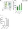
CSK overexpression increases TCR membrane localization (a, b). J76 cells transduced with DMF5 TCR and the indicated constructs were stained using MART-1 multimer. The mean fluorescence intensity (MFI) was plotted and each cell population was split into CSK–GFP positive and negative (dark green and light green, respectively) to compare cells from the same tube. a GFP vs. MART-1 multimer staining in CSK–GFP-transduced cells; and b one experiment representative of two separate experiments. c Sorted J76 cells expressing DMF5 TCR together with the indicated construct were stained with CD3 antibody and the MFI was plotted. This is representative of two separate experiments
Fig. 5
Fig. 5
CSK overexpression does not affect trogocytosis, but increases TCR-dependent target cell attachment. a T2 cells were loaded or not with a relevant (TGFβR2p) or an irrelevant peptide (M1p) O/N prior to co-incubation with J76 cells constitutively expressing Radium-1 TCR in combination with GFP (control), CSK–GFP or mCSK–GFP. The presence of surface CD3 was then monitored on T2 cells and the geometric mean plotted. b Same as in a but GFP+ cells (J76) were gated and the presence of HLA-A2 detected. Mean ± SD, n = 2. *P < 0.05, paired t test. c T2 cells used for a and b were stained with anti-HLA-A2 antibodies and MFI was plotted. Mean ± SD, n = 2. d T cells were electroporated with mRNA encoding for Radium-1 TCR and either GFP or e the mCSK–GFP. SCT expressing HeLa cells were used as APCs and were co-incubated with T cells for 15 min prior to live cell imaging. A total of 20 pictures per co-culture were taken; 10 pictures before washing (data not shown) and 10 pictures after washing the plate. GFP+ T cells bound to APCs were counted per frame. The dots represent the number of counts per frame after washing. Mean ± SD, n = 10. **P < 0.02, paired t test
Fig. 6
Fig. 6
Dummy T cells in a clinical setting. Design of the proposed procedure: patient T cells are isolated by leukapheresis and transfected with the therapeutic TCR and mCSK, preferably as mRNA. These cells are subsequently labelled with a bio-detectable agent and infused back into the patient. Accumulation of the signal will finally be monitored and exclusive homing to the tumour site or homing to additional sites will give a go/no go information to validate further use of the TCR for ACT

References

    1. Comrie WA, Burkhardt JK. Action and traction: cytoskeletal control of receptor triggering at the immunological synapse. Front Immunol. 2016;7:68. doi: 10.3389/fimmu.2016.00068. - DOI - PMC - PubMed
    1. Werlen G, Palmer E. The T-cell receptor signalosome: a dynamic structure with expanding complexity. Curr Opin Immunol. 2002;14(3):299–305. doi: 10.1016/S0952-7915(02)00339-4. - DOI - PubMed
    1. Malissen B, Bongrand P. Early T cell activation: integrating biochemical, structural, and biophysical cues. Annu Rev Immunol. 2015;33:539–561. doi: 10.1146/annurev-immunol-032414-112158. - DOI - PubMed
    1. Chakraborty AK, Weiss A. Insights into the initiation of TCR signaling. Nat Immunol. 2014;15(9):798–807. doi: 10.1038/ni.2940. - DOI - PMC - PubMed
    1. Chen L, Flies DB. Molecular mechanisms of T cell co-stimulation and co-inhibition. Nat Rev Immunol. 2013;13(4):227–242. doi: 10.1038/nri3405. - DOI - PMC - PubMed

MeSH terms

LinkOut - more resources