. 2018 Jan:27:27-39.
doi: 10.1016/j.ebiom.2017.12.004. Epub 2017 Dec 7.

T Cells Primed by Live Mycobacteria Versus a Tuberculosis Subunit Vaccine Exhibit Distinct Functional Properties

Affiliations

T Cells Primed by Live Mycobacteria Versus a Tuberculosis Subunit Vaccine Exhibit Distinct Functional Properties

Thomas Lindenstrøm et al. EBioMedicine. 2018 Jan.

Abstract

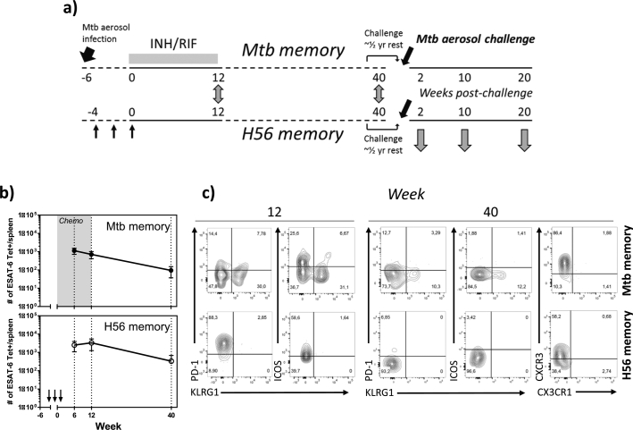
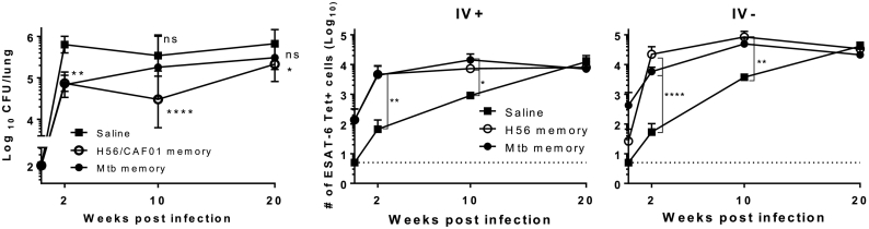
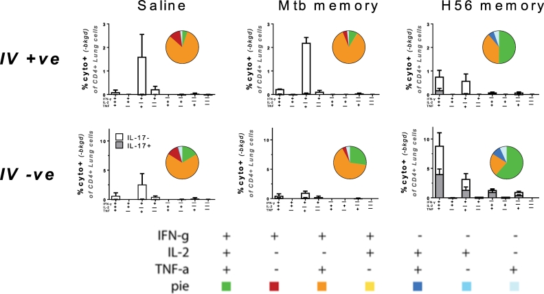
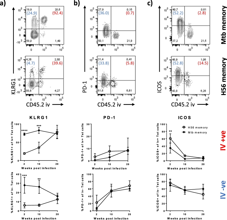
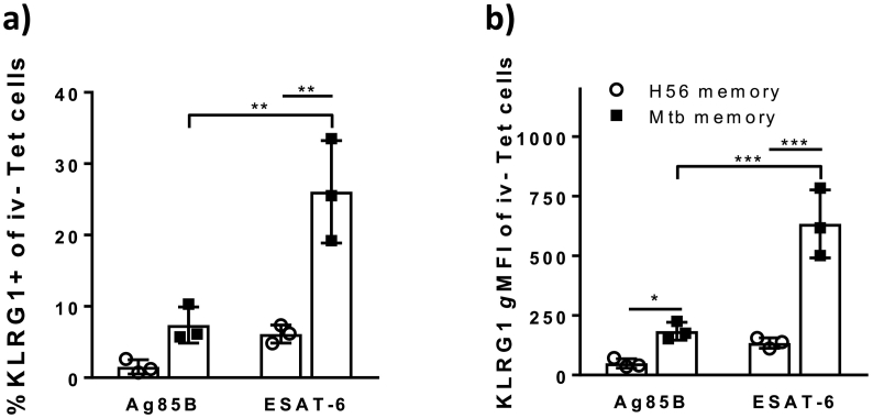
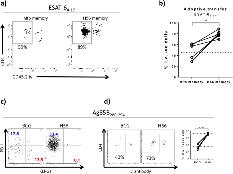
Despite inducing strong T cell responses, Mycobacterium tuberculosis (Mtb) infection fails to elicit protective immune memory. As such latently infected or successfully treated Tuberculosis (TB) patients are not protected against recurrent disease. Here, using a mouse model of aerosol Mtb infection, we show that memory immunity to H56/CAF01 subunit vaccination conferred sustained protection in contrast to the transient natural immunity conferred by Mtb infection. Loss of protection to re-infection in natural Mtb memory was temporally linked to an accelerated differentiation of ESAT-6- and to a lesser extent, Ag85B-specific CD4 T cells in both the lung parenchyma and vasculature. This phenotype was characterized by high KLRG1 expression and low, dual production of IFN-γ and TNF. In contrast, H56/CAF01 vaccination elicited cells that expressed low levels of KLRG1 with copious expression of IL-2 and IL-17A. Co-adoptive transfer studies revealed that H56/CAF01 induced memory CD4 T cells efficiently homed into the lung parenchyma of mice chronically infected with Mtb. In comparison, natural Mtb infection- and BCG vaccine-induced memory CD4 T cells exhibited a poor ability to home into the lung parenchyma. These studies suggest that impaired lung migratory capacity is an inherent trait of the terminally differentiated memory responses primed by mycobacteria/mycobacterial vectors.

Keywords: Lung homing; M. tuberculosis; Natural immunity; T cell priming & differentiation; Vaccination.


Figures

Fig. 1
Fig. 1
Resting memory cells after a cleared Mycobacterium tuberculosis infection show a higher degree of differentiation than memory cells primed by H56/CAF01 vaccination. A) Schematic overview of the experimental outline. C57BL/6 mice, aerosol infected for 6 weeks, were subjected to a 12 week Isoniacid/Rifabutin (INH/RIF) treatment. ESAT-6 specific memory cells in the spleen were phenotypically characterized at week 12/End of Treatment (EoT) by ESAT-64-17:I-Ab pulldown using magnetic bead-based tetramer enrichment and compared to E6 memory responses 12 week post three s.c. immunizations with H56/CAF01, each spaced by two weeks. ESAT-6 specific memory cells in the spleen were similarly characterized and compared between the two groups of resting memory mice at week 40 after immunization/start of treatment. In order to study protective efficacy, H56 and Mtb memory mice were allowed to rest for half a year and then aerosol infected with Mtb Erdman. Protective efficacy was evaluated at week 2, 10 and 20 after challenge. B) The number of ESAT-64-17-specific CD4 T cells in the spleen was determined by Tetramer pulldown in Mtb (upper panel) and H56 (lower panel) memory mice at week 6, 12 and 40. Symbols, mean ± s.d. of 3–4 mice per time point. For Mtb memory mice, the experiment was repeated twice at week 6 and once at week 12 and 40. For H56 mice, the experiment was repeated twice at week 6 and 12 and once for week 40. C) Representative flow cytometry plots depicting KLRG1, PD-1, ICOS and CXCR3 (wk 40) expression by ESAT-6 tetramer-binding CD4 T cells in the spleen of Mtb (upper panels) and H56 (lower panels) memory mice at week 12 and 40.
Fig. 2
Fig. 2
Mtb memory provides early, but transient, protection to Mycobacterium tuberculosis infection, whereas H56 memory mediates sustained protection accompanied by accelerated recruitment of E6-specific cells into the lung parenchyma. C57BL/6 mice were rested for ~ half a year prior to aerosol Mtb challenge (~ 100 CFU/lung). A) Total lung CFU was determined by serial plating at week 2, 10 and 20 in saline controls (black square), H56 memory (white circle) and Mtb memory (black circle). Data show mean ± s.d. of 8–14 mice/time-point in Saline group, and 6–7 mice/time-point in H56 and Mtb memory – though 4 at week 2 in these two groups. Two way ANOVA with Dunnett's multiple comparison test against Saline group. *P < 0.05; **P < 0.01, ****P < 0.0001. ns non-significant. B) Total number of ESAT-64–17 tetramer binding CD4 T cells within the lung vasculature (IV +) as determined by i.v. staining with anti-CD45 (unperfused lungs). C) Total number of lung parenchymal (IV-) ESAT-6-specific CD4 T cells (unperfused lungs). 3–4 mice/time-point. For B) & C): Cells gated on Singlets > Lymphocytes > Live, non-CD8s (Dead CD8) > CD3+ CD4+ > E6 Tet+ CD44+. For each time point, One-way ANOVA on log-transformed cell numbers with Tukey's multiple comparison test. *P < 0.05; **P < 0.01, ****P < 0.0001. A repeat experiment with similar overall outcome is shown in Supplementary Fig. 6.
Fig. 3
Fig. 3
Secondary effectors among memory mice differ in cytokine multifunctionality with responses in H56 memory mice dominated by IL-2- and IL-17A-producing CD4 T cell subsets. Ten weeks into Mtb challenge, lung cells from memory mice were i.v. stained with anti-CD45 prior to euthanization and subsequently stimulated ex vivo with ESAT-61-15 and stained by ICS to determine the frequency of antigen-specific CD4 T cells expressing IFN-g, TNF-a, IL-2 or IL-17A in any combination based on combinatorial Boolean gating analysis. Upper panels show lung vascular associated responses (IV +); lower panels responses in the lung parenchyma (IV −). Bar charts show mean frequencies ± s.d. (n = 3), white bars: IL-17A-; grey bars: IL-17A co-producers. Pies embedded into bar charts denote the proportion of each cytokine-producing subset of the responding cells from each lung compartment (unperfused lungs). Cells gated on Singlets > Lymphocytes > CD4 + > Combinatorial/Boolean gating on IV +, IFN-g +, TNF +, IL-2 +, IL-17A +. Repeated once with similar results.
Fig. 4
Fig. 4
ESAT-6-specific effectors in Mtb memory mice exhibit increased differentiation within the lung parenchyma early during infection compared to H56 memory mice. A) KLRG1 expression among lung localized I-Ab:ESAT-6-specific CD4 T cells. Representative FACS plots showing KLRG1 expression on ESAT-64-17 tet + cells from Mtb (upper plot) and H56 (lower plot) memory mice infected for ten weeks with Mycobacterium tuberculosis relative to their localization in the lung vasculature (CD45.2 IV + ve) or lung parenchyma (CD45.2 IV –ve). Numbers in parentheses represent the percentage of cells expressing KLRG1 in the IV + ve (Red) and the IV − ve population (Blue). Graphs show the proportion of KLRG1 + of I-Ab:ESAT-64-17 tetramer + CD4 T cells in the lung vasculature (IV + ve; upper graph) and in the lung parenchyma (IV –ve, lower graph) over the course of infection (unperfused lungs). Mean ± s.d. of 3–4 mice per group at any given time-point. Two-way ANOVA with Sidak's multiple comparison test for simple row effects. Differences between Mtb and H56 memory. ****P < 0.0001, ***P < 0.001, **P < 0.01. B) PD-1 expression among lung localized I-Ab:ESAT-6-specific CD4 T cells. Representative FACS plots showing PD-1 expression on ESAT-64–17 tet + cells from Mtb (upper plot) and H56 (lower plot) memory mice infected for ten weeks with Mycobacterium tuberculosis relative to their localization in the lung vasculature (CD45.2 IV + ve) or lung parenchyma (CD45.2 IV –ve). Numbers in parentheses represent the percentage of cells expressing PD-1 in the IV + ve (Red) and the IV − ve population (Blue). Graphs show the proportion of PD-1 + of I-Ab:ESAT-64-17 tetramer + CD4 T cells in the lung vasculature (IV + ve; upper graph) and in the lung parenchyma (IV –ve, lower graph) over the course of infection (unperfused lungs). Mean ± s.d. of 3–4 mice per group at any given time-point. Two-way ANOVA with Sidak's multiple comparison test for simple row effects (ns). C) ICOS expression among lung localized I-Ab:ESAT-6-specific CD4 T cells. Representative FACS plots showing ICOS expression on ESAT-64-17 tet + cells from Mtb (upper plot) and H56 (lower plot) memory mice infected for ten weeks with Mycobacterium tuberculosis relative to their localization in the lung vasculature (CD45.2 IV + ve) or lung parenchyma (CD45.2 IV –ve). Numbers in parentheses represent the percentage of cells expressing ICOS in the IV + ve (Red) and the IV –ve population (Blue). Graphs show the proportion of ICOS + of I-Ab:ESAT-64–17 tetramer + CD4 T cells in the lung vasculature (IV + ve; upper graph) and in the lung parenchyma (IV –ve, lower graph) over the course of infection (unperfused lungs). Mean ± s.d. of 3–4 mice per group at any given time-point. Two-way ANOVA with Sidak's multiple comparison test for simple row effects. Differences between Mtb and H56 memory. **P < 0.01. Gating as depicted in Supplementary Fig. 4. One of two comparable experiments shown.
Fig. 5
Fig. 5
ESAT-6 specific secondary effectors express significantly more KLRG1 than Ag85B-specific CD4 T cells in Mtb memory mice in contrast to H56 memory mice. Ten weeks into infection of rested C57BL/6 memory mice, the frequency of KLRG1 + cells and the expression levels of KLRG1 within the lung parenchyma (IV –ve population) was assessed after i.v. injection of anti-CD45.2-FITC prior to tissue harvest (unperfused lungs) using I-Ab:Ag85B280–294 and I-Ab:ESAT-64–17 specific tetramer staining. A) Bar chart showing the frequency of Ag85B280–294 and ESAT-64–17 specific CD4 T cells being KLRG1 + within the IV –ve lung population ten weeks into challenge of H56 (white) and Mtb memory (black) mice. Two-way ANOVA with Tukey's multiple comparison test. **P < 0.01. B) Bar chart showing the KLRG1 gMFIs of Ag85B280–294 and ESAT-64-17 specific CD4 T cells within the lung parenchyma (IV –ve) ten weeks into challenge of H56 (white) and Mtb memory (black) mice. Two-way ANOVA with Tukey's multiple comparison test. ***P < 0.001, *P < 0.05. Gating as depicted in Supplementary Fig. 4. The experiment was repeated once with similar results.
Fig. 6
Fig. 6
Antigen-specific CD4 T cells primed by mycobacteria are less efficient at migrating into the infected lung parenchyma than cells primed by H56/CAF01 vaccination in co-adoptive transfer experiments. A) CD4 T cells from long term H56 and Mtb memory mice were isolated from spleens and draining lymph nodes (inguinal and tracheobronchial, respectively) by negative selection and differentially stained with CPD450 (H56 memory) and CPD670 (Mtb memory) cell trackers, admixed in a ~ 1:1 ratio and adoptively transferred into recipient mice chronically infected with Mtb for 34 weeks. Location of I-Ab:ESAT-64-17-specific cells from each donor origin was assessed 16–18 h post transfer using tetramer-specific pulldown following i.v. administration of anti-CD45 just prior to euthanasia (unperfused lungs). Representative FACS plots showing ESAT-6 specific lung cells after enrichment using I-Ab:ESAT-64-17-specific tetramer pulldown. The two plots show the distribution in the lung compartment of ESAT-64-17 specific cells originating from Mtb memory donors (left) and H56 memory donors (right). The gate shows the percentage of ESAT-64-17 specific cells of Mtb memory vs H56 memory origin within the infected lung parenchyma (iv –ve). Gating as depicted in Supplementary Fig. 5. B) Graph showing the percentage of ESAT-64-17 specific cells of Mtb memory vs H56 memory donor origin within the I.V. –ve compartment (parenchyma) when co-adoptively transferred into the same infected recipients. Dotted lines depict mean values. Student's t-test, paired **P < 0.01. C) Congenic C57BL/6 mice were immunized with either 1 × BCG (CD45.1) or 3 × H56/CAF01 (CD45.2) and CD4 T cells purified by negative selection at ~ 50 days post vaccination. BCG and H56 memory cells were mixed in a1:1 ratio and Ag85B-sepcific responses characterized by I-Ab: Ag85B280–294 tetramer staining. Ag85B280–294 specific H56 (CD45.2) and BCG (CD45.1) memory cells were phenotypically characterized in terms of PD-1 and KLRG1 expression. No KLRG1 expressing Ag85B-specific cells were detected among H56 memory cells (left plot), which were dominated by PD-1 expression (blue). In contrast, a notable proportion of Ag85B280–294-specific cells primed by BCG (right plot) was found to express KLRG1 (red). D) Memory cells from BCG (CD45.1) and H56 (CD45.2) vaccinated mice were co-adoptively transferred into congenically-mismatched recipients carrying a 10 week old Mtb infection. Location of I-Ab: Ag85B280–294-specific cells from each donor origin was assessed based on their congenic markers (CD45.1 vs CD45.2) 20 h post transfer using tetramer-specific pulldown assays after i.v. labeling. Representative FACS plots showing the distribution of Ag85B280–294-specific memory cells primed by either BCG or H56/CAF01 within the lung of infected recipient mice. Graph summarizes the percentage of Ag85B280–294-specific memory cells primed by either BCG or H56/CAF01 within the parenchyma (I.V. – compartment) after co-adoptive transfer into the same recipients. Dotted lines depict mean values. Student's t-test, ****P < 0.0001. C & D – one representative experiment out of three repeat experiments shown.

References

    1. Aagaard C., Hoang T.T., Izzo A., Billeskov R., Troudt J., Arnett K., Keyser A., Elvang T., Andersen P., Dietrich J. Protection and polyfunctional T cells induced by Ag85B-TB10.4/IC31 against Mycobacterium tuberculosis is highly dependent on the antigen dose. PLoS One. 2009;4:e5930. - PMC - PubMed
    1. Aagaard C., Hoang T., Dietrich J., Cardona P.J., Izzo A., Dolganov G., Schoolnik G.K., Cassidy J.P., Billeskov R., Andersen P. A multistage tuberculosis vaccine that confers efficient protection before and after exposure. Nat. Med. 2011;17:189–U224. - PubMed
    1. Andersen P., Smedegaard B. CD4+ T-cell subsets that mediate immunological memory to Mycobacterium tuberculosis infection in mice. Infect. Immun. 2000;68:621–629. - PMC - PubMed
    1. Au-Yeung B.B., Smith G.A., Mueller J.L., Heyn C.S., Jaszczak R.G., Weiss A., Zikherman J. IL-2 modulates the TCR signaling threshold for CD8 but not CD4 T cell proliferation on a single-cell level. J. Immunol. 2017;198:2445–2456. - PMC - PubMed
    1. Baaten B.J., Cooper A.M., Swain S.L., Bradley L.M. Location, location, location: the impact of migratory heterogeneity on T cell function. Front. Immunol. 2013;4:311. - PMC - PubMed

MeSH terms