. 2018 Jan-Dec:31:394632017748841.
doi: 10.1177/0394632017748841. Epub 2017 Dec 18.

Ultrastructure of neurovascular changes in human diabetic retinopathy

Affiliations

Ultrastructure of neurovascular changes in human diabetic retinopathy

János Fehér et al. Int J Immunopathol Pharmacol. 2018 Jan-Dec.

Abstract

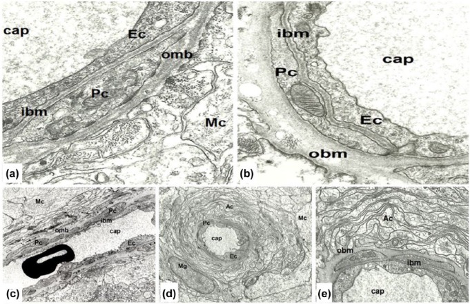
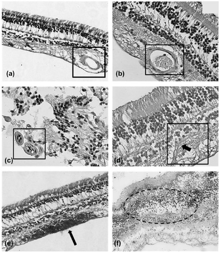
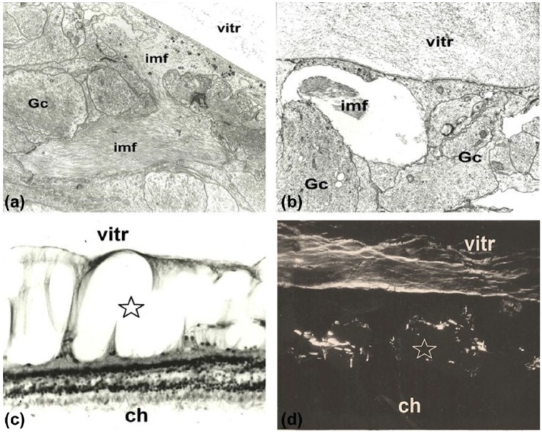
The previous concept regarding diabetic retinopathy assigned a primary role to hyperglycemia-induced microvascular alterations, while neuronal and glial abnormalities were considered to be secondary to either ischemia or exudation. The aim of this study was to reveal the potential role of neuronal and glial cells in initial and advanced alterations of the retinopathy in human type 2 diabetes. Electron microscopy and histochemical studies were performed on 38 surgically removed human eyes (28 obtained from diabetic patients and 10 from non-diabetic patients). Morphometric analysis of basement membrane material and lipids was performed. An accumulation of metabolic by-products was found in the capillary wall with aging: this aspect was significantly more pronounced in diabetics. Müller glial cells were found to contribute to alterations of the capillary wall and to occlusion, as well as to the development of proliferative retinopathy and cystoid degeneration of the retina. Our results showed morphological evidence regarding the role of neuronal and glial cells in the pathology of diabetic retinopathy, prior and in addition to microangiopathy. These morphological findings support a neurovascular pathogenesis at the origin of diabetic retinopathy, thus the current treatment approach should be completed by neuroprotective measures.

Keywords: basement membrane; diabetic macular edema; diabetic retinopathy; glia; proliferative diabetic retinopathy.


Conflict of interest statement

Declaration of conflicting interests: The author(s) declared no potential conflicts of interest with respect to the research, authorship, and/or publication of this article.

Figures

Figure 1.
Figure 1.
(a and b) Comparison of young and aged retinal capillary wall. (a) Retinal capillary (cap) in young age contains endothelium (Ec), pericyte (Pc), and Müller glial cells (Mc), and the basement membrane is formed by an inner leaflet (ibm) and an outer leaflet (obm). Six years male, magnification: ×24,000. (b) Aged retinal capillary (cap) contains an endothelial cell (Ec), pericyte (Pc), and significantly thickened basement membrane in which several electron-translucent, round-form vacuoles, and highly electron-dense granules are embedded. Both basement membrane thickening and lipid deposition affect the outer leaflet (obm) of the basement membrane, while the inner leaflet (ibm) is unaffected. Male 62 years, magnification ×24,000. (c–e) Retinal capillary in mild diabetes. (c) Retinal capillary (cap) in nearly longitudinal section: endothelial cells (Ec), pericytes (Pc), and their basement membrane (the inner leaflet of the capillary wall) show normal ultrastructural features. Müller cells (Mc) contain numerous, well-preserved organelles, and their basement membrane (the outer leaflet of the capillary wall) is significantly thickened; 81 years female, magnification: ×8000. (d) Capillary in the inner plexiform layer is surrounded by numerous cytoplasmic processes of astrocytes (Ac), microglia (Mg), and Müller glial cells (Mc) located around the “astrocyte-sheet.” Male 48 years, magnification ×8000. (e) Detail of the former picture in higher magnification. The inner leaflet of the basement membrane (ibm) is significantly thinner than the outer leaflet of the basement membrane (obm), which in several places extends in-between astrocytes. Endothelial cells and pericytes, as well as their basement membrane show normal appearance. Male 48 years, magnification ×20,000.
Figure 2.
Figure 2.
(a and b) Normal human retina. H&E stain. The normal morphology of the retina appears preserved in control patients. The lumen of the capillaries is free from glial cells proliferation, magnification ×40. (c and d) Human retina of patients affected by diabetic retinopathy. H&E stain. The lumen of the capillaries is filled by intravascular glial cells proliferation. In (c), two occluded capillaries in transverse section are visible (lower left corner). In (d), a capillary (longitudinal section) is occluded by intravascular glial cells’ proliferation (arrow), magnification ×40. (e and f) Human retina of patients affected by proliferative diabetic retinopathy. H&E stain. Within the retinal tissue, some microhemorrhages (e, arrow) and cystoid degeneration of the inner layers of the retina (f) are visible, magnification ×20.
Figure 3.
Figure 3.
Cystoid retinal degeneration. (a) Early phase of cystoid retinal degeneration: accumulation of intermediate filaments (imf) between glial cells (Gc) is visible. Male 40 years, transmission electron microscopy. Magnification ×16,000. (b) Early cystoid degeneration of the retina with intermediate filaments (imf) in one of the empty spaces between glial cells (Gc). No neuronal elements may be identified. In the vitreous (vitr), disorganized filaments and fine granular material are visible. Female 56 years. Transmission electron microscopy. Magnification ×18,000. (c) Advanced cystoid degeneration of the retina (star). Some columns of Müller cells separate empty “cystoid” spaces of the inner retina, while outer layers are well-preserved. Note the increased thickness of the retina (ch: choroid, vitr: vitreous). Female 69 years. Light microscopy: H&E stain. Magnification ×800. 3D. Glycoprotein-rich filaments are visible in the cystoid spaces of the retina (star) and in the vitreous body (ch: choroid, vitr: vitreous). Female 69 years. Polarization microscopic picture. Magnification ×800.

Similar articles

Cited by

References

    1. Barber AJ. (2015) Diabetic retinopathy: Recent advances towards understanding neurodegeneration and vision loss. Science China Life Sciences 58(6): 541–549. - PubMed
    1. Jonsson KB, Frydkjaer-Olsen U, Grauslund J. (2016) Vascular changes and neurodegeneration in the early stages of diabetic retinopathy: Which comes first? Ophthalmic Research 56(1): 1–9. - PubMed
    1. Sohn EH, Van Dijk HW, Jiao C, et al. (2016) Retinal neurodegeneration may precede microvascular changes characteristic of diabetic retinopathy in diabetes mellitus. Proceedings of the National Academy of Sciences of the United States of America 113(19): E2655–E2664. - PMC - PubMed
    1. Lynch SK, Abràmoff MD. (2017) Diabetic retinopathy is a neurodegenerative disorder. Vision Research. Epub ahead of print 28 April DOI: 10.1016/j.visres.2017.03.003. - DOI - PMC - PubMed
    1. Coughlin BA, Feenstra DJ, Mohr S. (2017) Müller cells and diabetic retinopathy. Vision Research. Epub ahead of print 5 September DOI: 10.1016/j.visres.2017.03.013. - DOI - PMC - PubMed

LinkOut - more resources