Review
. 2017 Dec 1;109(20):1680-1710.
doi: 10.1002/bdr2.1180.

Blood-brain barrier development: Systems modeling and predictive toxicology

Affiliations
Review

Blood-brain barrier development: Systems modeling and predictive toxicology

Katerine S Saili et al. Birth Defects Res. .

Abstract

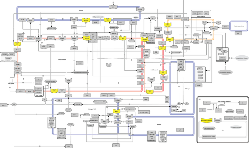
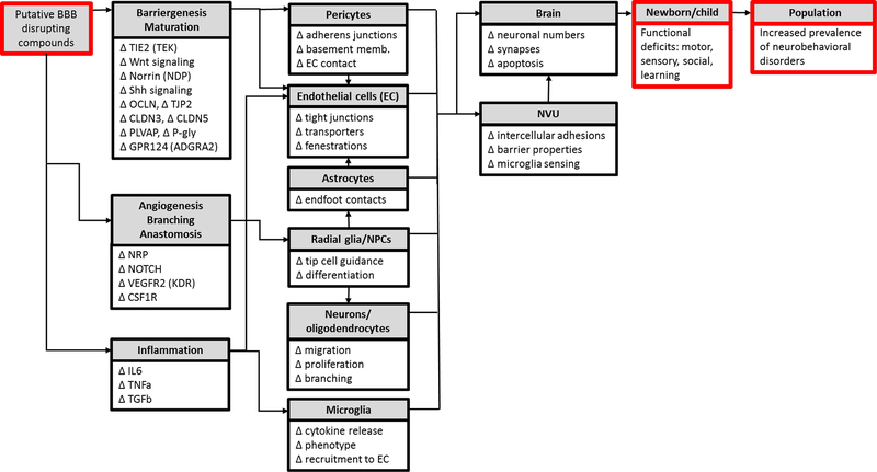
The blood-brain barrier (BBB) serves as a gateway for passage of drugs, chemicals, nutrients, metabolites, and hormones between vascular and neural compartments in the brain. Here, we review BBB development with regard to the microphysiology of the neurovascular unit (NVU) and the impact of BBB disruption on brain development. Our focus is on modeling these complex systems. Extant in silico models are available as tools to predict the probability of drug/chemical passage across the BBB; in vitro platforms for high-throughput screening and high-content imaging provide novel data streams for profiling chemical-biological interactions; and engineered human cell-based microphysiological systems provide empirical models with which to investigate the dynamics of NVU function. Computational models are needed that bring together kinetic and dynamic aspects of NVU function across gestation and under various physiological and toxicological scenarios. This integration will inform adverse outcome pathways to reduce uncertainty in translating in vitro data and in silico models for use in risk assessments that aim to protect neurodevelopmental health.

Keywords: blood-brain barrier; brain; children's environmental health; embryo; neurovascular development; neurovascular unit; predictive toxicology.


Conflict of interest statement

6. Conflict of interest statement

The authors declare no conflicts.

Figures

Figure 1:
Figure 1:. Neurovascular unit (NVU).
A) NVU showing endothelial cells (red), pericytes (light purple), astrocytes (bright purple), neurons (dark purple), and microglia (orange); color coded to match Figure 3. B) Neural tube, perineural vascular plexus (PNVP), neural crest, and neural ectoderm. Transverse section at mouse Thieler Stage (TS) 16 (E10).
Figure 2:
Figure 2:. Phylogeny of the BBB.
Primary amino acid sequence similarity for 86 proteins implicated in BBB development (see figure 4) was determined for species representing classes that appear at different times throughout evolutionary history: (Mammalia: Homo sapiens, Pan troglodytes, Mus musculus; Actinopterygii (ray-finned fish): Danio rerio; Chondrichthyes (cartilaginous fish): Callorhinchus milii; Leptocardii (earliest vertebrates): Branchiostoma belcheri; Branchiopoda (crustaceans): Daphnia magna; Insecta: Musca domestica; Hyperoartia (invertebrate chordate): Lethenteron camtschaticum; Cephalopoda: Eteroctopus dofleini). Comparisons to the human protein (100%) were made by an amino acid sequence query using the Basic Local Alignment Search Tool for proteins (BLASTp) of the National Center for Biotechnology Information (NCBI) protein database using SeqAPASS v2.0 software (https://seqapass.epa.gov/seqapass) (LaLone et al., 2016), which facilitates consistent comparisons of amino acid sequence similarity across multiple species. For proteins with multiple isoforms, SeqAPASS best practices were used to select the most representative NCBI protein accession number for the BLAST; and full sequence lengths (rather than functional domains) were used. A beta testing version of ToxPi software v2.0 provided by David Reif (NCSU) was used to produce the sequence similarity ToxPis using the ‘single’ clustering algorithm (Reif et al., 2010). For each protein, the % similarity to the human protein (100%) is represented by the length of pie slice (i.e., shorter pie slices represent lower similarity; or absence of a slice indicates no sequence similarity). Slices are arranged from highest to lowest % similarity in the mouse compared to humans (i.e., red slices have the highest similarity and light purple slices represent the lowest similarity). The type of BBB (glial or endothelial) is indicated on the dendrogram at the node representing a common ancestor with the same type of BBB. The symbols listed in the ‘protein’ key correspond to standard NCBI gene names. NCBI protein database accessed between 6/27/17 and 7/27/17.
Figure 3:
Figure 3:. Timeline of NVU development.
The boxed area indicates mouse Thieler Stages (TS) 9 (E6.5) to 27 (newborn). The section above the box roughly indicates the corresponding human Carnegie Stages (CS) and gestational days (GD) beginning at human CS 6–7 (GD14 −16). Neurons, oligodendrocytes, astrocytes, and forebrain pericytes derive from neural ectoderm (purple); while endothelial cells, mid- and hindbrain pericytes, and microglia derive from mesoderm (red). Vasculogenesis begins during mouse TS 12 (E8.5), angiogenesis/invasion occurs at TS 15 (E9.5), lateral branching and tight junctions are present by TS 16 (E10), and anastomosis and microglia peak at TS 19 (E11.5).
Figure 4:
Figure 4:. Control network for neurovascular development.
Biowiring diagram with representative signaling pathways, receptors, and ligands that are important for blood-brain barrier (BBB) development. Large rectangles are cells (color-coded to match Figures 1 and 2): stalk and tip endothelial cells (red); pericyte, radial glia/NPC, neuron, astrocyte, head mesenchyme (purple); microglia (orange). Yellow highlights represent key receptors hypothesized to mediate developmental NVU toxicity. Molecule abbreviations are standard human NCBI gene symbols (see Table 1 for names).
Figure 5:
Figure 5:. Preliminary AOP framework for BBB development and function.
An adverse outcome pathway (AOP) for BBB development should include the proposed molecular initiating events (MIEs), key events (KEs), and adverse outcomes (AOs) resulting from embryonic or fetal exposure to a putative BBB disrupting compound. The list of potential MIEs/KEs included here encompasses molecules important for BBB development (summarized in Figure 4). Perturbation of these molecules is indicated by ∆ to indicate that any change (either an increase or decrease in expression) could potentially lead to BBB disruption. These molecular events underlie the function of the cells of the NVU. Altered cellular function is expected to impact NVU and/or brain development and function, leading to adverse outcomes such as neurodevelopmental toxicity and neurobehavioral disorders.

References

    1. Abbott NJ, 2013. Blood-brain barrier structure and function and the challenges for CNS drug delivery. J Inherit Metab Dis 36(3), 437–449. - PubMed
    1. Abbott NJ, Lane NJ, Bundgaard M, 1992. A fibre matrix model for the restricting junction of the blood-brain barrier in a cephalopod mollusc: implications for capillary and epithelial permeability. J Neurocytol 21(4), 304–311. - PubMed
    1. Abbott NJ, Patabendige AA, Dolman DE, Yusof SR, Begley DJ, 2010. Structure and function of the blood-brain barrier. Neurobiol Dis 37(1), 13–25. - PubMed
    1. Achyuta AK, Conway AJ, Crouse RB, Bannister EC, Lee RN, Katnik CP, Behensky AA, Cuevas J, Sundaram SS, 2013. A modular approach to create a neurovascular unit-on-a-chip. Lab Chip 13(4), 542–553. - PubMed
    1. Adriani G, Ma D, Pavesi A, Kamm RD, Goh EL, 2016. A 3D neurovascular microfluidic model consisting of neurons, astrocytes and cerebral endothelial cells as a blood-brain barrier. Lab Chip 17(3), 448–459. - PubMed