. 2018 Jan 1;28(1):1-8.
doi: 10.1093/cercor/bhw342.

Inhibitory Modulation of Orbitofrontal Cortex on Medial Prefrontal Cortex-Amygdala Information Flow

Affiliations

Inhibitory Modulation of Orbitofrontal Cortex on Medial Prefrontal Cortex-Amygdala Information Flow

Chun-Hui Chang et al. Cereb Cortex. .

Abstract

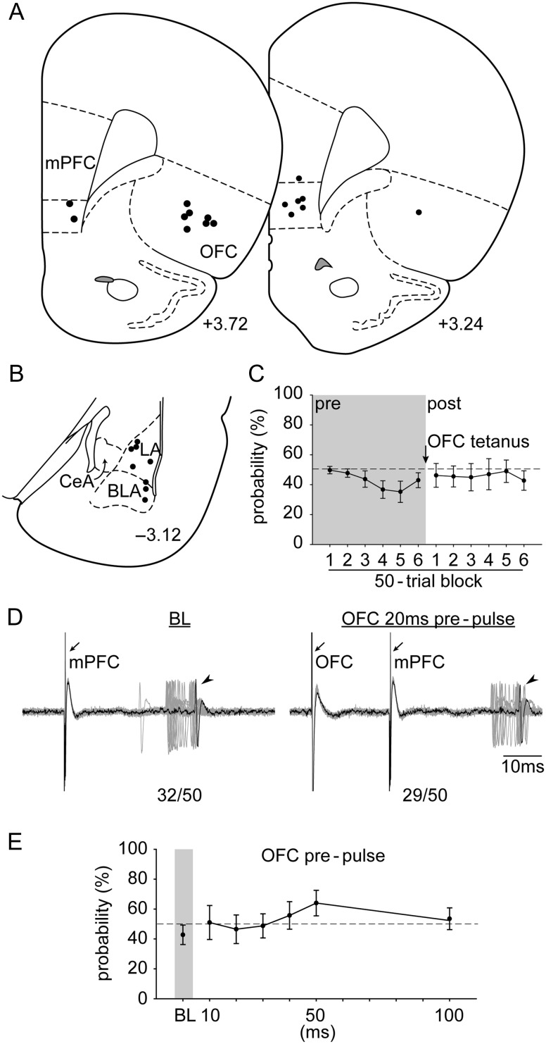
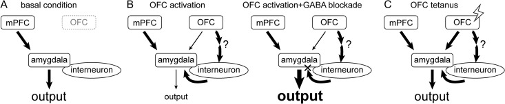
The amygdala receives cortical inputs from the medial prefrontal cortex (mPFC) and orbitofrontal cortex (OFC) that are believed to affect emotional control and cue-outcome contingencies, respectively. Although mPFC impact on the amygdala has been studied, how the OFC modulates mPFC-amygdala information flow, specifically the infralimbic (IL) division of mPFC, is largely unknown. In this study, combined in vivo extracellular single-unit recordings and pharmacological manipulations were used in anesthetized rats to examine how OFC modulates amygdala neurons responsive to mPFC activation. Compared with basal condition, pharmacological (N-Methyl-D-aspartate) or electrical activation of the OFC exerted an inhibitory modulation of the mPFC-amygdala pathway, which was reversed with intra-amygdala blockade of GABAergic receptors with combined GABAA and GABAB antagonists (bicuculline and saclofen). Moreover, potentiation of the OFC-related pathways resulted in a loss of OFC control over the mPFC-amygdala pathway. These results show that the OFC potently inhibits mPFC drive of the amygdala in a GABA-dependent manner; but with extended OFC pathway activation this modulation is lost. Our results provide a circuit-level basis for this interaction at the level of the amygdala, which would be critical in understanding the normal and pathophysiological control of emotion and contingency associations regulating behavior.

Keywords: amygdala; in vivo electrophysiology; medial prefrontal cortex; orbitofrontal cortex; rat.


Figures

Figure 1.
Figure 1.
Representative electrical stimulation sites and cannula placement in (A) the mPFC and OFC, as well as a representative recording site in (B) the LA (+3.72, and −3.00; AP distance [mm] to bregma). Open arrowheads, the lesion marks at the tips of the stimulation electrodes. Closed arrowhead, tip of the infusion cannula. Arrow, dye mark of the neuron recorded. LA, lateral nucleus of the amygdala; BLA, basolateral nucleus of the amygdala; CeA, central nucleus of the amygdala.
Figure 2.
Figure 2.
The placements of (A) all the stimulation electrodes in the mPFC and infusion cannulae in the OFC and (B) the distribution of all the neurons recorded (+3.72, +3.24, −2.92, −3.12, and −3.36; AP distance [mm] to bregma). (C) Electrophysiological recording of an amygdala neuron responsive to mPFC stimulation (left) that decreased its evoked responses after OFC pharmacological activation with NMDA (right; n/100 = evoked spikes out of 100 trials). Arrows, electrical stimulation artifacts from mPFC stimulation. Arrowheads, evoked spikes in amygdala. (D) Pharmacological activation of the OFC with NMDA exerted an inhibitory modulation on the mPFC–amygdala pathway (*P < 0.05 relative to VEH and APV). VEH, vehicle. Other abbreviations refer to Figure 1.
Figure 3.
Figure 3.
The placements of (A) all the stimulation electrodes in the mPFC and the OFC and (B) the distribution of all the neurons recorded (+3.72, +3.24, −2.92, −3.12, and −3.36; AP distance [mm] to bregma). (C) Electrophysiological recording of an amygdala neuron responsive to mPFC stimulation (left) that showed a decrease in evoked responses with OFC 20 ms prepulse (n/50 = evoked spikes out of 50 trials). Arrows, electrical stimulation artifacts from OFC and mPFC stimulation, respectively. Arrowheads, evoked spikes in amygdala. (D) OFC activation exerted an inhibitory gating on the mPFC–amygdala pathway at all delays tested (relative to BL; *P < 0.05). Abbreviations refer to Figure 1.
Figure 4.
Figure 4.
The placements of (A) all the stimulation electrodes in the mPFC and the OFC and (B) the distribution of all the neurons recorded (+3.72, +3.24, and −3.12; AP distance [mm] to bregma). (C) Electrophysiological recording of an amygdala neuron that is responsive to mPFC stimulation that exhibited a decrease in evoked responses following OFC 20 ms prepulse. This was reversed to a facilitation upon local administration of GABA antagonists, followed by a rapid return to inhibitory gating (n/50 = evoked spikes out of 50 trials). (D) Compared with BL, evoked probability was significantly decreased with OFC prepulse (20 ms; “a”, P < 0.05), and significantly increased under the influence of GABA antagonists (“b”, P < 0.05). Abbreviations refer to Figures 1 and 3.
Figure 5.
Figure 5.
The placements of (A) all the stimulation electrodes in the mPFC and the OFC and (B) the distribution of all the neurons recorded (+3.72, +3.24, and −3.12; AP distance [mm] to bregma). (C) OFC tetanus did not change evoked probability of the amygdala neuron response to mPFC stimulation. (D) Electrophysiological recording of an amygdala neuron that responded to mPFC stimulation after OFC tetanus. (E) Stimulation of the OFC failed to produce an inhibitory gating over the mPFC–amygdala pathway following OFC tetanus. Abbreviations refer to Figures 1 and 3.
Figure 6.
Figure 6.
One potential model that may account for the OFC modulation of the mPFC–amygdala pathway. Compared with (A) basal condition, (B) pharmacological or electrical activation of the OFC exerted an inhibitory modulation of the mPFC–amygdala pathway (left), which was reversed by intra-amygdala blockade of GABAergic receptors (right). (C) Tetanization of the OFC-related pathways results in a loss of OFC control over the mPFC–amygdala pathway, presumably because of a selective enhancement of the OFC–amygdala principle neuron input. Abbreviations refer to Figure 1.

References

    1. Aggleton JP, Burton MJ, Passingham RE. 1980. Cortical and subcortical afferents to the amygdala of the rhesus monkey (Macaca mulatta). Brain Res. 190:347–368. - PubMed
    1. Asher A, Lodge DJ. 2012. Distinct prefrontal cortical regions negatively regulate evoked activity in nucleus accumbens subregions. Int J Neuropsychopharmacol. 15:1287–1294. - PMC - PubMed
    1. Buffalari DM, Grace AA. 2007. Noradrenergic modulation of basolateral amygdala neuronal activity: opposing influences of alpha-2 and beta receptor activation. J Neurosci. 27:12358–12366. - PMC - PubMed
    1. Canteras NS, Swanson LW. 1992. Projections of the ventral subiculum to the amygdala, septum, and hypothalamus: a PHAL anterograde tract-tracing study in the rat. J Comp Neurol. 324:180–194. - PubMed
    1. Chang CH, Grace AA. 2014. Amygdala-ventral pallidum pathway decreases dopamine activity after chronic mild stress in rats. Biol Psychiatry. 76:223–230. - PMC - PubMed

Publication types

MeSH terms