. 2018 Apr 15;197(8):1027-1035.
doi: 10.1164/rccm.201707-1474OC.

Novel Method for Noninvasive Sampling of the Distal Airspace in Acute Respiratory Distress Syndrome

Affiliations

Novel Method for Noninvasive Sampling of the Distal Airspace in Acute Respiratory Distress Syndrome

J Brennan McNeil et al. Am J Respir Crit Care Med. .

Abstract

Rationale: A major barrier to a more complete understanding of acute respiratory distress syndrome (ARDS) pathophysiology is the inability to sample the distal airspace of patients with ARDS. The heat moisture exchanger (HME) filter is an inline bacteriostatic sponge that collects exhaled moisture from the lungs of mechanically ventilated patients.

Objectives: To test the hypothesis that HME filter fluid (HMEF) represents the distal airspace fluid in patients with ARDS.

Methods: Samples of HMEF were collected from 37 patients with acute pulmonary edema (either from ARDS or hydrostatic causes [HYDRO; control subjects]). Concurrent undiluted pulmonary edema fluid (EF) and HMEF were collected from six patients. HMEF from 11 patients (8 ARDS and 3 HYDRO) were analyzed by liquid chromatography-coupled tandem mass spectometry. Total protein (bicinchoninic acid assay), MMP-9 (matrix metalloproteinase-9), and MPO (myeloperoxidase) (ELISA) were measured in 29 subjects with ARDS and 5 subjects with HYDRO. SP-D (surfactant protein-D), RAGE (receptor for advanced glycation end-products) (ELISA), and cytokines (IL-1β, IL-6, IL-8, and tumor necrosis factor-α) (electrochemiluminescent assays) were measured in six concurrent HMEF and EF samples.

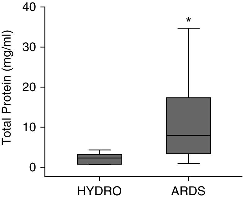
Measurements and main results: Liquid chromatography-coupled tandem mass spectrometry on concurrent EF and HMEF samples from four patients revealed similar base peak intensities and m/z values indicating similar protein composition. There were 21 significantly elevated proteins in HMEF from patients with ARDS versus HYDRO. Eight proteins measured in concurrent EF and HMEF from six patients were highly correlated. In HMEF, total protein and MMP-9 were significantly higher in ARDS than in HYDRO.

Conclusions: These data suggest that HMEF is a novel, noninvasive method to accurately sample the distal airspace in patients with ARDS.

Keywords: acute respiratory distress syndrome; airspace fluid; biomarkers; heat moisture exchanger filter; pulmonary edema fluid..


Figures

Figure 1.
Figure 1.
Depiction of a heat moisture exchanger (HME) filter placement inline in the ventilator circuit between the patient and mechanical ventilator.
Figure 2.
Figure 2.
Direct spectral comparison of matched heat moisture exchanger filter fluid (HMEF) and edema fluid (EF) samples. Visual comparison of matched pulmonary EF (upgoing peaks, blue) and HMEF (downgoing peaks, red) reveals similar base peak intensities and locations in a patient with acute pulmonary edema from acute respiratory distress syndrome (ARDS) (A) and one with hydrostatic pulmonary edema (HYDRO) (B). Bold peaks bearing a dot are reflected in spectra from both HMEF and EF samples. There were 118 common peaks in matched ARDS samples and 157 common peaks in matched HYDRO samples.
Figure 3.
Figure 3.
Correlation of spectral intensities between matched heat moisture exchanger (HME) filter fluid and edema fluid samples. Label-free quantitation of spectral intensities from four patients with matched HME filter fluid and edema fluid collection reveals correlation of spectral intensities within each patient (white squares, r2 range = 0.849–0.951) and less correlation between patients (gray squares, r2 range = 0.121–0.835). Patient 4, acute respiratory distress syndrome; Patient 2, hydrostatic pulmonary edema; Patients 1 and 3, mixed.
Figure 4.
Figure 4.
Total protein concentrations in matched edema fluid (EF) and heat moisture exchanger filter fluid (HMEF). (A) Total protein on matched EF (solid circles) and HMEF (open triangles) from six patients. Patients 1 and 3, acute respiratory distress syndrome; Patient 4, hydrostatic pulmonary edema; Patients 2, 5, and 6, mixed. (B) A Bland-Altman plot of agreement between HMEF and EF total protein (dashed lines represent upper and lower 95% confidence intervals). The diamonds show the difference between the measurements on each fluid for an individual patient.
Figure 5.
Figure 5.
Individual protein quantification of eight analytes in matched heat moisture exchanger filter fluid (HMEF) and edema fluid (EF). Individual proteins were measured by ELISA and electrochemiluminescence in six patients with matched samples of HMEF and EF. Patients 1 and 3, acute respiratory distress syndrome; Patient 4, hydrostatic pulmonary edema; Patients 2, 5, and 6, mixed. MMP-9 = matrix metalloproteinase-9; MPO = myeloperoxidase; RAGE = receptor for advanced glycation end-products; SP-D = surfactant protein-D; TNF-α = tumor necrosis factor-α.
Figure 6.
Figure 6.
Total protein comparison in heat moisture exchanger filter fluid between patients with acute respiratory distress syndrome (ARDS) and those with hydrostatic pulmonary edema (HYDRO). Total protein was higher in heat moisture exchanger filter fluid from patients with ARDS (n = 29) than in that from patients with HYDRO (n = 5). *P = 0.008. Box-and-whisker plots show median (line), interquartile range (IQR) (box ends), and 1.5 × IQR (whiskers).
Figure 7.
Figure 7.
MMP-9 (matrix metalloproteinase-9) and MPO (myeloperoxidase) quantification. Protein quantification of (A) MMP-9 and (B) MPO in heat moisture exchanger filter fluid from 29 patients with acute respiratory distress syndrome (ARDS) and 5 patients with hydrostatic pulmonary edema (HYDRO). *P = 0.022; MPO, P = 0.179. Box-and-whisker plots show median (line), interquartile range (IQR) (box ends), and 1.5 × IQR (whiskers).
Figure 8.
Figure 8.
Protein interactions in heat moisture exchanger filter fluid for patients with acute respiratory distress syndrome. High-confidence interactions between proteins elevated in heat moisture exchanger filter fluid demonstrate notable interactions between detected proteins. The green area highlights MMP-9, a previously described mediator of lung injury.

Comment in

References

    1. Pierrakos C, Vincent JL. The changing pattern of acute respiratory distress syndrome over time: a comparison of two periods. Eur Respir J. 2012;40:589–595. - PubMed
    1. Wang CY, Calfee CS, Paul DW, Janz DR, May AK, Zhuo H, et al. One-year mortality and predictors of death among hospital survivors of acute respiratory distress syndrome. Intensive Care Med. 2014;40:388–396. - PMC - PubMed
    1. Brower RG, Matthay MA, Morris A, Schoenfeld D, Thompson BT, Wheeler A Acute Respiratory Distress Syndrome Network. Ventilation with lower tidal volumes as compared with traditional tidal volumes for acute lung injury and the acute respiratory distress syndrome. N Engl J Med. 2000;342:1301–1308. - PubMed
    1. National Heart, Lung, and Blood Institute Acute Respiratory Distress Syndrome (ARDS) Clinical Trials Network, Wiedemann HP, Wheeler AP, Bernard GR, Thompson BT, Hayden D, et al. Comparison of two fluid-management strategies in acute lung injury. N Engl J Med. 2006;354:2564–2575. - PubMed
    1. Wattiez R, Falmagne P. Proteomics of bronchoalveolar lavage fluid. J Chromatogr B Analyt Technol Biomed Life Sci. 2005;815:169–178. - PubMed

Publication types

MeSH terms