. 2018 Mar;1863(3):284-298.
doi: 10.1016/j.bbalip.2017.12.005. Epub 2017 Dec 16.

Mitochondrial CDP-diacylglycerol synthase activity is due to the peripheral protein, TAMM41 and not due to the integral membrane protein, CDP-diacylglycerol synthase 1

Affiliations

Mitochondrial CDP-diacylglycerol synthase activity is due to the peripheral protein, TAMM41 and not due to the integral membrane protein, CDP-diacylglycerol synthase 1

Nicholas J Blunsom et al. Biochim Biophys Acta Mol Cell Biol Lipids. 2018 Mar.

Abstract

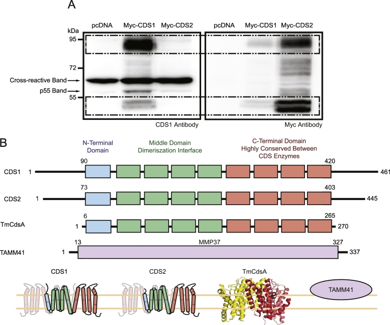
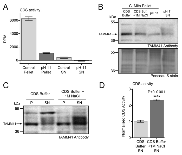
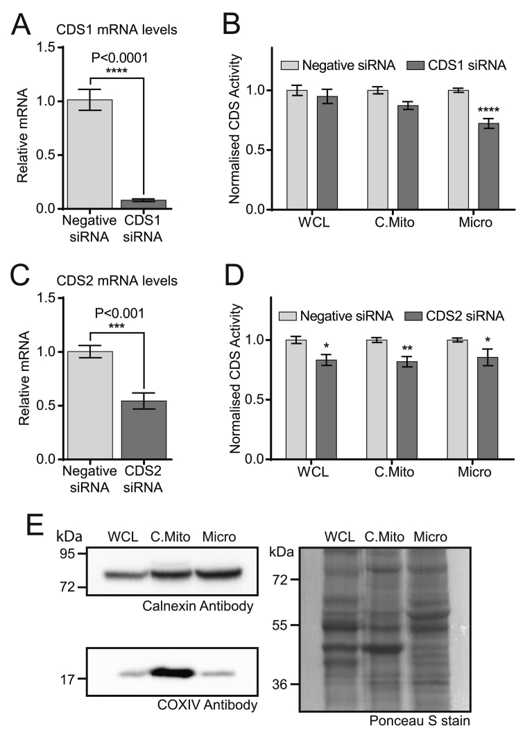
CDP diacylglycerol synthase (CDS) catalyses the conversion of phosphatidic acid (PA) to CDP-diacylglycerol, an essential intermediate in the synthesis of phosphatidylglycerol, cardiolipin and phosphatidylinositol (PI). CDS activity has been identified in mitochondria and endoplasmic reticulum of mammalian cells apparently encoded by two highly-related genes, CDS1 and CDS2. Cardiolipin is exclusively synthesised in mitochondria and recent studies in cardiomyocytes suggest that the peroxisome proliferator-activated receptor γ coactivator 1 (PGC-1α and β) serve as transcriptional regulators of mitochondrial biogenesis and up-regulate the transcription of the CDS1 gene. Here we have examined whether CDS1 is responsible for the mitochondrial CDS activity. We report that differentiation of H9c2 cells with retinoic acid towards cardiomyocytes is accompanied by increased expression of mitochondrial proteins, oxygen consumption, and expression of the PA/PI binding protein, PITPNC1, and CDS1 immunoreactivity. Both CDS1 immunoreactivity and CDS activity were found in mitochondria of H9c2 cells as well as in rat heart, liver and brain mitochondria. However, the CDS1 immunoreactivity was traced to a peripheral p55 cross-reactive mitochondrial protein and the mitochondrial CDS activity was due to a peripheral mitochondrial protein, TAMM41, not an integral membrane protein as expected for CDS1. TAMM41 is the mammalian equivalent of the recently identified yeast protein, Tam41. Knockdown of TAMM41 resulted in decreased mitochondrial CDS activity, decreased cardiolipin levels and a decrease in oxygen consumption. We conclude that the CDS activity present in mitochondria is mainly due to TAMM41, which is required for normal mitochondrial function.

Keywords: Cardiolipin; Differentiation; Heart; Mitochondria; PITPNC1; Phosphatidic acid; Retinoic acid.


Figures

None
Graphical abstract
Fig. 1
Fig. 1
Differentiation of H9c2 cells with retinoic acid. [A] Morphology examined by Nomarski optics (upper panels) and cellular f-actin and nuclei stained with phalloidin and DAPI respectively (lower panels). [B] Western blot of cell lysates (50 μg protein) obtained from proliferating cells (10% FCS) and differentiated cells in either 1% FCS or 1% FCS with 1 μM retinoic acid (RA). [C] Example traces of oxygen consumption in proliferating and differentiated cells (1% FCS with RA); arrows indicate addition of drugs, and maximal ETS (electron transport system) indicated by a dotted line (nmt, non-mitochondrial). [D] Quantitation of the Routine, Leak and maximal ETS in proliferating and differentiated cells. The results are averages from two independent experiments for the differentiated cells (1% FCS with RA) and from three experiments for the proliferating cells. Error bars denote ± S.E.M.
Fig. 2
Fig. 2
Localisation of CDS1 immunoreactivity and CDS activity to mitochondria. [A, B] Differentiated H9c2 cells fractionated and analysed for [A] CDS1 immunoreactivity by Western blot; [B] CDS activity. [C-E] Rat heart fractionated and analysed for [C] CDS1 immunoreactivity and markers, [D] CDS1 immunoreactivity (entire Western blot shown) and [E] CDS activity. [F–H] Rat heart fractionated in the presence of the protease, subtilisin and analysed for [F] CDS1 immunoreactivity by western blot, [G] CDS activity and [H] PI synthase (PIS) activity. CDS and PIS activity was monitored in triplicate and error bars denote ± S.E.M. COXIV, GRP75 and cyto c are markers for mitochondria, PITPNC1 and PITPα are cytosolic markers and calnexin is a marker for the ER. WCL, whole cell lysate; Micro, microsomes; C.Mito, crude mitochondria; Cyto, cytosol; P.Mito, pure mitochondria, MAMs, mitochondrial associated membranes; PIS, PI synthase; CDS, CDP-diacylglycerol synthase.
Fig. 3
Fig. 3
CDS1 immunoreactivity and CDS activity localises to mitochondria in rat brain and liver. [A, C] Rat brain and [B, D] liver fractionated and analysed by CDS1 immunoreactivity by Western blot and for CDS activity. CDS activity was monitored in triplicate and error bars denote S.E.M. WCL, whole cell lysate; Micro, microsomes; C.Mito, crude mitochondria; Cyto, cytosol; P.Mito, pure mitochondria, MAMs, mitochondrial associated membranes; CDS, CDP-diacylglycerol synthase.
Fig. 4
Fig. 4
Differentiation of H9c2 cells does not cause an increase in CDS1 mRNA expression. H9c2 cells were differentiated with retinoic acid and samples removed daily to monitor mRNA levels. [A] CDS1 mRNA; [B] CDS2 mRNA; [C] TAMM41 mRNA; [D] PITPNC1 mRNA. Data from 3 experiments done in triplicate ± S.E.M.
Fig. 5
Fig. 5
CDS1 antibody recognises over-expressed CDS1 but not over-expressed CDS2. [A] Myc-tagged CDS1 and CDS2 were expressed in COS-7 cells and membranes obtained after centrifugation. Membranes (25 μg protein per lane) probed with antibodies to CDS1 (left panel) and Myc (right panel). The boxed region highlights the over-expressed proteins. CDS proteins run as monomers (~ 45 kDa) and as dimers (~ 95 kDa). The CDS1 antibody (Abcam) also recognises a p55 protein in the CDS1-over-expressing cells. [B] Topological organisation of the CDS enzymes, CDS1, CDS2 and TmCdsA based on the structure of TmCdsA, and the structurally unrelated CDS enzyme, TAMM41. The transmembrane domains of CDS1 and CDS2 are shown in coloured boxes. Blue boxes, N-terminal domain, green boxes, middle domain which is the dimerization interface and the orange boxes which is the highly-conserved C-terminal domain. Also shown is the three dimensional structure of TmCdsA which forms a dimer; the monomers are coloured red or yellow. Cartoon representation of CDS1 and CDS2 based on TmCdsA dimer. TAMM41 (NP_001271330.1) is shown as a peripheral membrane protein containing the MMP37 domain (MMP37, mitochondrial matrix proteins of 37 kDa; PFAM: 09139) .
Fig. 6
Fig. 6
Knockdown of CDS1 with siRNA does not lead to a reduction in the p55 band. [A] Detection of the p55 band with the CDS1 antibody in undifferentiated H9c2 cells is only possible when mitochondrial fractions are prepared. [B] Validation of CDS1 siRNA. A decrease in CDS1 mRNA (P < 0.0001, n = 6, ± S.E.M.) but not CDS2 or TAMM41 is observed when undifferentiated H9c2 cells were treated with CDS1 siRNA. [C] Mitochondrial fractions prepared from control, negative siRNA-treated and CDS1 siRNA-treated H9c2 cells were analysed for CDS1 immunoreactivity by Western blot. WCL, whole cell lysate; C.Mito, crude mitochondria; Micro, microsomes.
Fig. 7
Fig. 7
Mitochondrial CDS activity is due to a peripheral membrane protein. [A, B] Total membranes (50 μg protein) prepared from COS-7 cells expressing either Myc-CDS1 or Myc-CDS2 were analysed by western blot [A] before and [B] after treatment with 0.2 M sodium bicarbonate (pH 11). [C] Loading Control: Ponceau S stain of blots in [A]. [D] CDS activity of COS-7 cell total membranes (50 μg protein) before and after treatment with sodium bicarbonate. [E, F] Mitochondria purified from rat brain were incubated with high salt (1 M NaCl) or with sodium bicarbonate to remove peripheral proteins. [E] The residual mitochondrial membranes and supernatant were analysed by Western blot with CDS1 and COXIV antibody. [F] The membranes were analysed for CDS activity.
Fig. 8
Fig. 8
Mitochondrial CDS activity is due to TAMM41 and its knockdown reduces oxygen consumption. [A] Knockdown of TAMM1 mRNA by siRNA in H9c2 cells (n = 3, unpaired two-tailed t-test). [B] Western blot showing knockdown of TAMM41 protein in the crude mitochondrial fractions from TAMM41 knockdown cells. Loading control: Ponceau S stain of the blot. Crude mitochondria were prepared from H9c2 cells treated with control or TAMM41 siRNA. Representative blot of two independent crude mitochondrial fractions. [C] Quantification of TAMM41 protein in crude mitochondria from 4 independent fractionations (n = 4, unpaired two-tailed t-test). [D] CDS activity after TAMM41 knockdown in H9c2 subcellular fractions. Results are from two independent experiments done in triplicate (n = 6, two-way ANOVA with multiple comparisons). The average activity in control whole cell lysates from different experiments was in the range of 2–6 pmol/mg/min. WCL, whole cell lysate; C.Mito, crude mitochondria; Micro, microsomes; [E] Cardiolipin levels in crude mitochondria prepared from control and TAMM41 siRNA-treated H9c2 cells. H9c2 cells were labelled to near equilibrium with 14C-acetate for 72 h and lipids extracted from crude mitochondrial fractions and analysed by TLC. Results are from 3 independent experiments done in duplicate or triplicate (n = 8, unpaired two-tailed t-test). [F] Example trace of oxygen consumption in control and TAMM41 knockdown H9c2 cells; arrows indicate addition of drugs and maximal ETS (electron transport system) shown by dotted line. nmt, non-mitochondrial; O, oligomycin; F, FCCP; A, antimycin A. [G] Quantitation of the Routine, Leak and maximal ETS in control and TAMM41-knockdown cells. The results are averages from three independent experiments; two way ANOVA with multiple comparisons Error bars denote ± S.E.M.
Fig. 9
Fig. 9
TAMM41 protein is released from the mitochondria after treatment with high salt and its activity is recovered in the supernatant. [A] Liver mitochondria were incubated with CDS buffer (50 mM Tris-HCL (pH 8.0), 50 mM KCl, 0.2 mM EGTA) or with 0.2 M sodium bicarbonate (pH 11). The supernatants were recovered, concentrated and buffer exchanged. CDS activity was monitored in the pellets and the supernatants. [B] Western blot of crude mitochondria treated with 1 M NaCl and pH 11 buffer and pH 11 supernatant. [C] Liver mitochondria were incubated with control buffer or control buffer supplemented with 1 M sodium chloride for 1 h. The mitochondrial pellets and the respective supernatants were Western blotted with TAMM41 antibody. P., pellet; SN, supernatant. [D] The supernatant from [C] was concentrated, buffer exchanged and CDS activity was assessed. Data from two experiments performed in triplicate ± S.E.M.
Fig. 10
Fig. 10
Crude mitochondria contain residual CDS1 and CDS2 activity that can be accounted for by ER associated membranes. [A, C] Knockdown of [A] CDS1 mRNA and [C] CDS2 mRNA by siRNA in H9c2 cells. [B, D] CDS activity after [B] CDS1 knockdown and [D] CDS2 knockdown in H9c2 subcellular fractions. Results from three independent experiments in triplicate were combined. In each experiment, the control sample was set at 1 and the knockdown samples are a proportion of the control. [E] Subcellular fractions (WCL, crude mitochondria and microsomes) analysed by Western blot for calnexin (ER marker) and COXIV (mitochondrial marker). A representative blot is shown from four separate fractionations.

References

    1. Mejia E.M., Nguyen H., Hatch G.M. Mammalian cardiolipin biosynthesis. Chem. Phys. Lipids. 2014;179:11–16. - PubMed
    1. Palmer J.W., Tandler B., Hoppel C.L. Biochemical properties of subsarcolemmal and interfibrillar mitochondria isolated from rat cardiac muscle. J. Biol. Chem. 1977;252:8731–8739. - PubMed
    1. Hom J., Sheu S.S. Morphological dynamics of mitochondria—a special emphasis on cardiac muscle cells. J. Mol. Cell. Cardiol. 2009;46:811–820. - PMC - PubMed
    1. Scarpulla R.C., Vega R.B., Kelly D.P. Transcriptional integration of mitochondrial biogenesis. Trends Endocrinol. Metab. 2012;23:459–466. - PMC - PubMed
    1. Hock M.B., Kralli A. Transcriptional control of mitochondrial biogenesis and function. Annu. Rev. Physiol. 2009;71:177–203. - PubMed

Publication types

MeSH terms

Substances