. 2017 Dec 18;13(12):e1007132.
doi: 10.1371/journal.pgen.1007132. eCollection 2017 Dec.

Leaf shedding as an anti-bacterial defense in Arabidopsis cauline leaves

Affiliations

Leaf shedding as an anti-bacterial defense in Arabidopsis cauline leaves

O Rahul Patharkar et al. PLoS Genet. .

Abstract

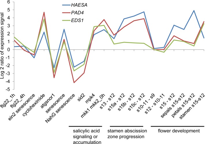
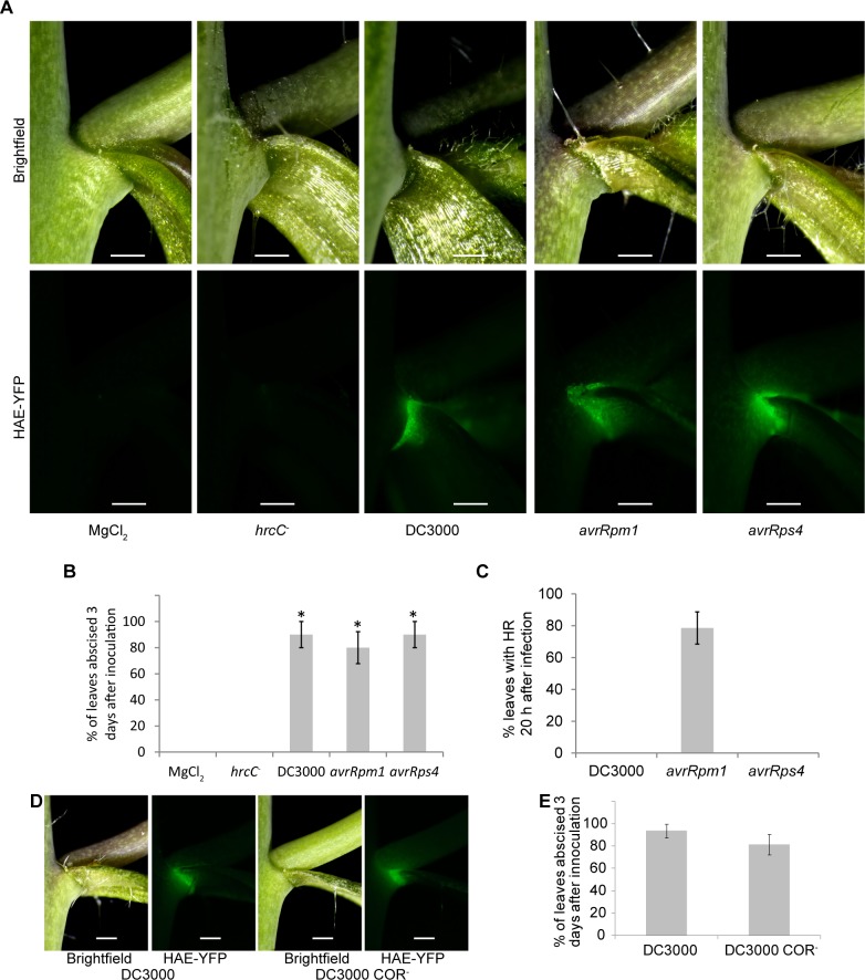
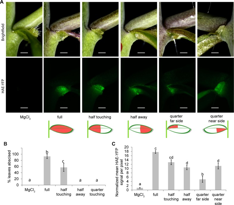
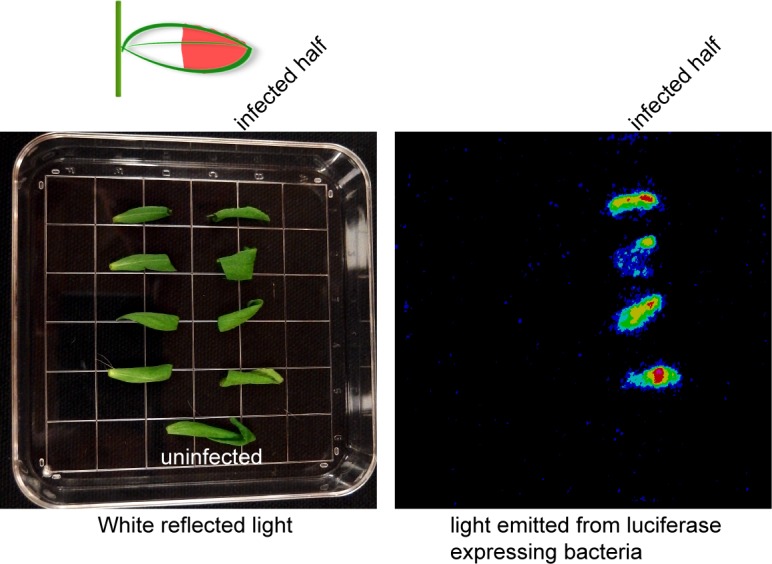
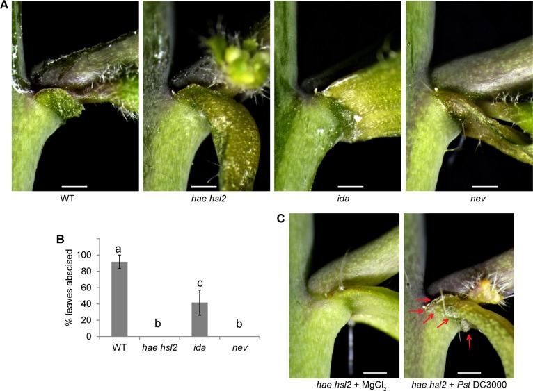
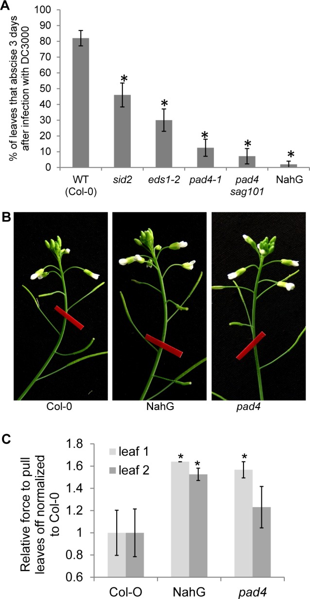
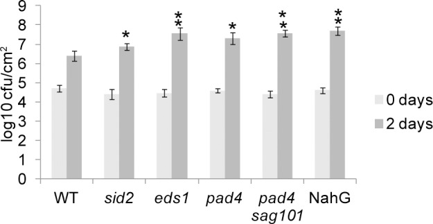
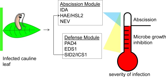
Plants utilize an innate immune system to protect themselves from disease. While many molecular components of plant innate immunity resemble the innate immunity of animals, plants also have evolved a number of truly unique defense mechanisms, particularly at the physiological level. Plant's flexible developmental program allows them the unique ability to simply produce new organs as needed, affording them the ability to replace damaged organs. Here we develop a system to study pathogen-triggered leaf abscission in Arabidopsis. Cauline leaves infected with the bacterial pathogen Pseudomonas syringae abscise as part of the defense mechanism. Pseudomonas syringae lacking a functional type III secretion system fail to elicit an abscission response, suggesting that the abscission response is a novel form of immunity triggered by effectors. HAESA/HAESA-like 2, INFLORESCENCE DEFICIENT IN ABSCISSION, and NEVERSHED are all required for pathogen-triggered abscission to occur. Additionally phytoalexin deficient 4, enhanced disease susceptibility 1, salicylic acid induction-deficient 2, and senescence-associated gene 101 plants with mutations in genes necessary for bacterial defense and salicylic acid signaling, and NahG transgenic plants with low levels of salicylic acid fail to abscise cauline leaves normally. Bacteria that physically contact abscission zones trigger a strong abscission response; however, long-distance signals are also sent from distal infected tissue to the abscission zone, alerting the abscission zone of looming danger. We propose a threshold model regulating cauline leaf defense where minor infections are handled by limiting bacterial growth, but when an infection is deemed out of control, cauline leaves are shed. Together with previous results, our findings suggest that salicylic acid may regulate both pathogen- and drought-triggered leaf abscission.


Conflict of interest statement

The authors have declared that no competing interests exist.

Figures

Fig 1
Fig 1. HAESA is co-expressed with PAD4 and EDS1 in a number of different tissues and treatments.
Publicly available microarray data indicates that PAD4 and EDS1 are statistically increased during the abscission process in stamen abscission zones [27]. Furthermore, HAE, PAD4, and EDS1 expression are up in shoot and leaf tissues of mutants with increased SA levels and down in mutants with decreased SA levels [55,56,31,57,58].
Fig 2
Fig 2. Bacteria with a viable type III secretion system can activate HAE expression and trigger cauline leaf abscission.
(A) Plants expressing HAE-YFP (driven by the HAE promoter) were infected with virulent or avirulent DC3000. Images are 2 days after infiltration. The same samples are shown in the top and bottom panels where the top panels are imaged with reflected white light and the bottom panels are imaging YFP fluorescence. (B) Abscission of cauline leaves three days after infection. Data are mean ± s.e.m.; n = 5 biological replicates (one plant each); t-test versus MgCl2 control; *P < 0.005. (C) Hypersensitive response in cauline leaves 20 h after infection. Data are mean ± s.e.m.; n = 7 biological replicates (one plant each, 2 leaves per plant). (D) Leaves infected with DC3000 or DC3000 that does not produce coronatine (COR-) for two days. (E) Abscission of cauline leaves 3 days after infection. Data are mean ± s.e.m.; n = 8 biological replicates (one plant each). Scale bar is 0.5 mm.
Fig 3
Fig 3. Abscission occurs when DC3000 touches the AZ, however, long-distance signals are also sent from distal portions of the leaf to the AZ.
(A) Leaves infiltrated in indicated portions of the leaf shown 3 days after infection. The same samples are shown in the top and bottom panels where the top panels are imaged with reflected white light and the bottom panels are imaging YFP fluorescence. (B) Percent of cauline leaves to abscise three days after treatment. Data are mean ± s.e.m.; n = 7 biological replicates (one plant each); letters indicated different statistical quantities t-test P < 0.05. (C) Quantification of HAE-YFP fluorescence two days after infection while all leaves were still attached. Data are mean ± s.e.m.; n = 4 biological replicates (one plant each); letters indicate different statistical quantities t-test P < 0.05. Scale bar is 0.5 mm.
Fig 4
Fig 4. DC3000 does not move in cauline leaves from the place of infiltration.
The indicated half of each cauline leaf was infiltrated with DC3000 that express luciferase constitutively driven by the kanamycin promoter. A line was drawn on the leaf with a pen to indicate the border of the infiltrated/not infiltrated. The leaves were cut in half 2 days after infection and imaged with white reflected light (left) and luminescence (right). Four replicates were performed with the same results.
Fig 5
Fig 5. The floral organ abscission pathway is necessary for pathogen-triggered leaf abscission.
(A) Photos of cauline leaf AZ of WT plants and floral abscission defective mutants treated with DC3000 for 3 days. (B) Percent cauline leaves abscised 3 days after infection. Data are mean ± s.e.m.; n = 6 biological replicates (one plant each); letters indicated different statistical quantities t-test P < 0.05. (C) Micrograph of cauline leaf AZs from hae hsl2 plants treated with or without DC3000. Images are representative from at least 4 replicates. Red arrows indicate enlarged AZ cells. Scale bar is 0.5 mm.
Fig 6
Fig 6. Pathogen defense defective mutants are defective in pathogen-triggered cauline leaf abscission.
(A) Percent of cauline leaves that abscised 3 days after infection. Data are mean ± s.e.m.; n = 25 biological replicates (one plant each); t-test versus WT; *P < 0.0001. (B) NahG and pad4 floral organ abscission is similar to WT. Red tape is 9.5 mm wide. (C) Relative leaf breakstrength force of WT (Col-0), NahG transgenic plants, and pad4 exposed to drought and re-watering conditions. Data are mean ± s.e.m.; n = 12 biological replicates (one plant each); t-test versus WT; *P < 0.05.
Fig 7
Fig 7. Bacterial growth in cauline leaves of Arabidopsis defense mutants.
Bacterial enumeration in cauline leaf 2. Data are mean ± s.e.m.; n = 5 biological replicates (one plant each); t-test versus WT; *P < 0.1, **P < 0.05.
Fig 8
Fig 8. Proposed cauline leaf defense model.
Cauline leaf microbial defense has a threshold system. Minor infections are fought to limit the multiplication of bacterial growth. Microbial growth inhibition requires PAD4, EDS1, SAG101, and SID2 (and salicylic acid). If the infection becomes too serious, the entire cauline leaf will simply be abscised. Abscission requires the previously mentioned defense components as well as HAE/HSL2, IDA, and NEV. Yellow and blue triangles symbolize that when infection becomes severe the defense module and abscission module have overlapping function. The defense module potentially regulates the abscission module via salicylic acid where salicylic acid induces expression of HAE.

References

    1. Abramovitch RB, Anderson JC, Martin GB. Bacterial elicitation and evasion of plant innate immunity. Nat Rev Mol Cell Biol. 2006;7: 601–611. doi: 10.1038/nrm1984 - DOI - PMC - PubMed
    1. Jones JDG, Dangl JL. The plant immune system. Nat Lond. 2006;444: 323–9. http://dx.doi.org/10.1038/nature05286 - DOI - PubMed
    1. Zipfel C. Pattern-recognition receptors in plant innate immunity. Curr Opin Immunol. 2008;20: 10–16. doi: 10.1016/j.coi.2007.11.003 - DOI - PubMed
    1. Melotto M, Underwood W, He SY. Role of Stomata in Plant Innate Immunity and Foliar Bacterial Diseases. Annu Rev Phytopathol. 2008;46: 101–122. doi: 10.1146/annurev.phyto.121107.104959 - DOI - PMC - PubMed
    1. Dodds PN, Rathjen JP. Plant immunity: towards an integrated view of plant–pathogen interactions. Nat Rev Genet. 2010;11: 539–548. doi: 10.1038/nrg2812 - DOI - PubMed

Publication types