

Cerebrospinal fluid metabolomic profiles can discriminate patients with leptomeningeal carcinomatosis from patients at high risk for leptomeningeal metastasis
- PMID: 29254157
- PMCID: PMC5731867
- DOI: 10.18632/oncotarget.20983
Cerebrospinal fluid metabolomic profiles can discriminate patients with leptomeningeal carcinomatosis from patients at high risk for leptomeningeal metastasis
Abstract
Purpose: Early diagnosis of leptomeningeal carcinomatosis (LMC) is necessary to improve outcomes of this formidable disease. However, cerebrospinal fluid (CSF) cytology is frequently false negative. We examined whether CSF metabolome profiles can be used to differentiate patients with LMC from patients having a risk for development of LMC.
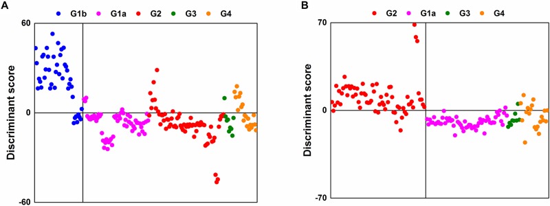
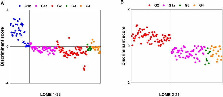
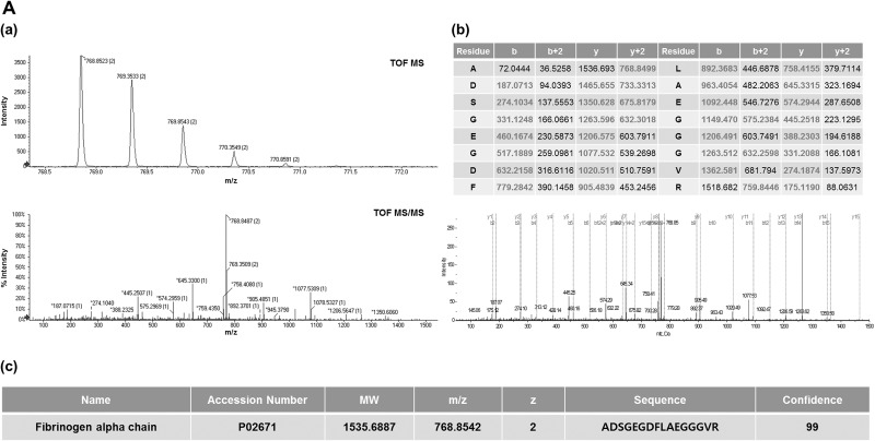
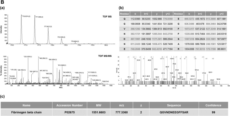
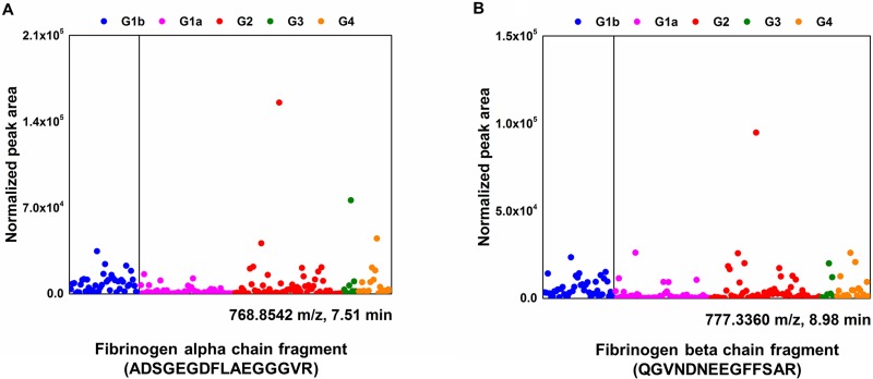
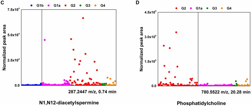
Results: A total of 10,905 LMIs were evaluated using PCA-DA. The LMIs defined Group 2 with a sensitivity of 85% and a specificity of 91%. After selecting 33 LMIs, including diacetylspermine and fibrinogen fragments, the CSF metabolomics profile had a sensitivity of 100% and a specificity of 93% for discriminating Group 1b from the other groups. After selecting 21 LMIs, including phosphatidylcholine, the CSF metabolomics profile differentiated LMC (Group 2) patients from the high-risk groups of Group 3 and Group 4 with 100% sensitivity and 100% specificity.
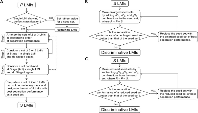
Materials and methods: We prospectively collected CSF from five groups of patients: Group 1a, systemic cancer; Group 1b, no tumor; Group 2, LMC; Group 3, brain metastasis; Group 4, brain tumor other than brain metastasis. All metabolites in the CSF samples were detected as low-mass ions (LMIs) using mass spectrometry. Principal component analysis-based discriminant analysis (PCA-DA) and two search algorithms were used to select the LMIs that differentiated the patient groups of interest from controls.
Conclusions: Analysis of CSF metabolite profiles could be used to diagnose LMC and exclude patients at high-risk of LMC with a 100% accuracy. We expect a future validation trial to evaluate CSF metabolic profiles supporting CSF cytology.
Keywords: cerebrospinal fluid; diagnosis; leptomeningeal carcinomatosis; metabolome; profile.
Conflict of interest statement
CONFLICTS OF INTEREST None.
Figures
References
-
- Debus OM, Lerchl A, Bothe HW, Bremer J, Fiedler B, Franssen M, Koehring J, Steils M, Kurlemann G. Spontaneous central melatonin secretion and resorption kinetics of exogenous melatonin: a ventricular CSF study. J Pineal Res. 2002;33:213–7. - PubMed
-
- Frankfort SV, Tulner LR, van Campen JP, Verbeek MM, Jansen RW, Beijnen JH. Amyloid beta protein and tau in cerebrospinal fluid and plasma as biomarkers for dementia: a review of recent literature. Curr Clin Pharmacol. 2008;3:123–31. - PubMed
-
- Romeo MJ, Espina V, Lowenthal M, Espina BH, Petricoin EF, 3rd, Liotta LA. CSF proteome: a protein repository for potential biomarker identification. Expert Rev Proteomics. 2005;2:57–70. - PubMed
-
- Diez B, Balmaceda C, Matsutani M, Weiner HL. Germ cell tumors of the CNS in children: recent advances in therapy. Childs Nerv Syst. 1999;15:578–85. - PubMed
-
- Nakagawa H, Kubo S, Murasawa A, Nakajima S, Nakajima Y, Izumoto S, Hayakawa T. Measurements of CSF biochemical tumor markers in patients with meningeal carcinomatosis and brain tumors. J Neurooncol. 1992;12:111–20. - PubMed
LinkOut - more resources
Full Text Sources
Other Literature Sources
Molecular Biology Databases
