

New reconstruction algorithm for digital breast tomosynthesis: better image quality for humans and computers
- PMID: 29254355
- PMCID: PMC6088454
- DOI: 10.1177/0284185117748487
New reconstruction algorithm for digital breast tomosynthesis: better image quality for humans and computers
Abstract
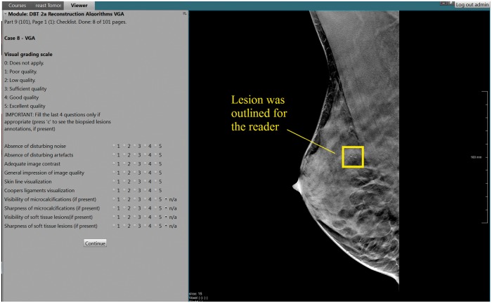
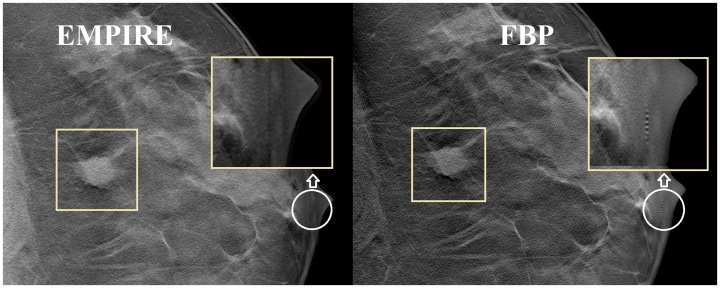
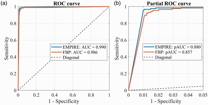
Background The image quality of digital breast tomosynthesis (DBT) volumes depends greatly on the reconstruction algorithm. Purpose To compare two DBT reconstruction algorithms used by the Siemens Mammomat Inspiration system, filtered back projection (FBP), and FBP with iterative optimizations (EMPIRE), using qualitative analysis by human readers and detection performance of machine learning algorithms. Material and Methods Visual grading analysis was performed by four readers specialized in breast imaging who scored 100 cases reconstructed with both algorithms (70 lesions). Scoring (5-point scale: 1 = poor to 5 = excellent quality) was performed on presence of noise and artifacts, visualization of skin-line and Cooper's ligaments, contrast, and image quality, and, when present, lesion visibility. In parallel, a three-dimensional deep-learning convolutional neural network (3D-CNN) was trained (n = 259 patients, 51 positives with BI-RADS 3, 4, or 5 calcifications) and tested (n = 46 patients, nine positives), separately with FBP and EMPIRE volumes, to discriminate between samples with and without calcifications. The partial area under the receiver operating characteristic curve (pAUC) of each 3D-CNN was used for comparison. Results EMPIRE reconstructions showed better contrast (3.23 vs. 3.10, P = 0.010), image quality (3.22 vs. 3.03, P < 0.001), visibility of calcifications (3.53 vs. 3.37, P = 0.053, significant for one reader), and fewer artifacts (3.26 vs. 2.97, P < 0.001). The 3D-CNN-EMPIRE had better performance than 3D-CNN-FBP (pAUC-EMPIRE = 0.880 vs. pAUC-FBP = 0.857; P < 0.001). Conclusion The new algorithm provides DBT volumes with better contrast and image quality, fewer artifacts, and improved visibility of calcifications for human observers, as well as improved detection performance with deep-learning algorithms.
Keywords: Digital breast tomosynthesis; deep learning; reconstruction algorithms; visual grading analysis.
Figures


References
-
- Tabar L, Gad A, Holmberg L, et al. Reduction in mortality from breast cancer after mass screening with mammography: randomised trial from the Breast Cancer Screening Working Group of the Swedish National Board of Health and Welfare. Lancet 1985; 325: 829–832. - PubMed
-
- Independent UK Panel on Breast Cancer Screening. The benefits and harms of breast cancer screening: an independent review. Lancet 2012; 380: 1778–1786. - PubMed
-
- Andersson I, Ikeda DM, Zackrisson S, et al. Breast tomosynthesis and digital mammography: a comparison of breast cancer visibility and BIRADS classification in a population of cancers with subtle mammographic findings. Eur Radiol 2008; 18: 2817–2825. - PubMed
-
- Skaane P, Gullien R, Bjorndal H, et al. Digital breast tomosynthesis (DBT): initial experience in a clinical setting. Acta Radiol 2012; 53: 524–529. - PubMed
MeSH terms
LinkOut - more resources
Full Text Sources
Other Literature Sources
Medical
Miscellaneous
