

Prismatic Adaptation Modulates Oscillatory EEG Correlates of Motor Preparation but Not Visual Attention in Healthy Participants
- PMID: 29255004
- PMCID: PMC5792477
- DOI: 10.1523/JNEUROSCI.1422-17.2017
Prismatic Adaptation Modulates Oscillatory EEG Correlates of Motor Preparation but Not Visual Attention in Healthy Participants
Abstract
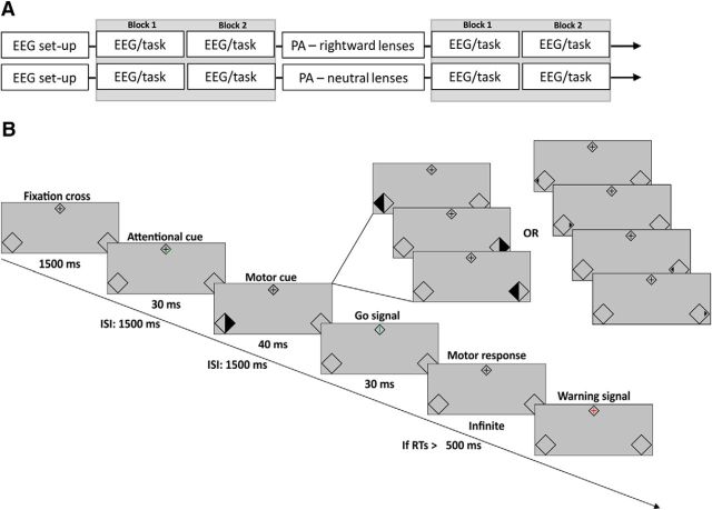
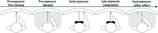
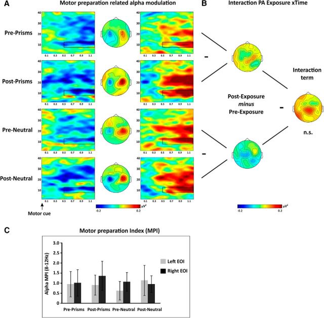
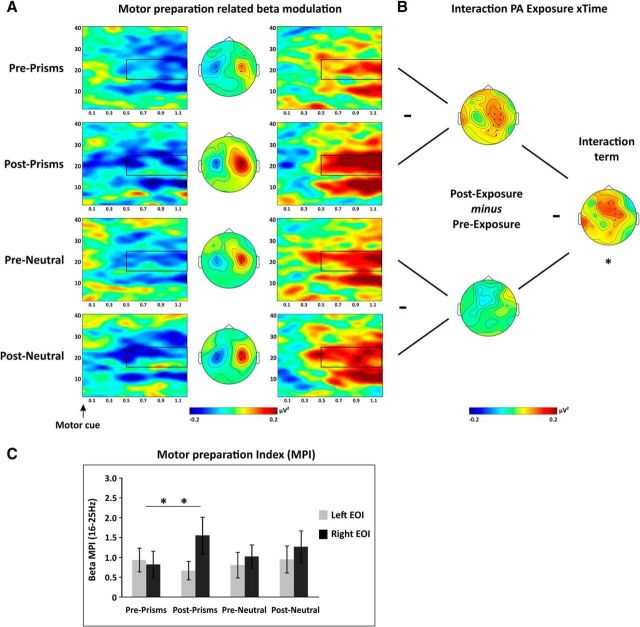
Prismatic adaption (PA) has been proposed as a tool to induce neural plasticity and is used to help neglect rehabilitation. It leads to a recalibration of visuomotor coordination during pointing as well as to aftereffects on a number of sensorimotor and attention tasks, but whether these effects originate at a motor or attentional level remains a matter of debate. Our aim was to further characterize PA aftereffects by using an approach that allows distinguishing between effects on attentional and motor processes. We recorded EEG in healthy human participants (9 females and 7 males) while performing a new double step, anticipatory attention/motor preparation paradigm before and after adaptation to rightward-shifting prisms, with neutral lenses as a control. We then examined PA aftereffects through changes in known oscillatory EEG signatures of spatial attention orienting and motor preparation in the alpha and beta frequency bands. Our results were twofold. First, we found PA to rightward-shifting prisms to selectively affect EEG signatures of motor but not attentional processes. More specifically, PA modulated preparatory motor EEG activity over central electrodes in the right hemisphere, contralateral to the PA-induced, compensatory leftward shift in pointing movements. No effects were found on EEG signatures of spatial attention orienting over occipitoparietal sites. Second, we found the PA effect on preparatory motor EEG activity to dominate in the beta frequency band. We conclude that changes to intentional visuomotor, rather than attentional visuospatial, processes underlie the PA aftereffect of rightward-deviating prisms in healthy participants.SIGNIFICANCE STATEMENT Prismatic adaptation (PA) has been proposed as a tool to induce neural plasticity in both healthy participants and patients, due to its aftereffect impacting on a number of visuospatial and visuomotor functions. However, the neural mechanisms underlying PA aftereffects are poorly understood as only little neuroimaging evidence is available. Here, we examined, for the first time, the origin of PA aftereffects studying oscillatory brain activity. Our results show a selective modulation of preparatory motor activity following PA in healthy participants but no effect on attention-related activity. This provides novel insight into the PA aftereffect in the healthy brain and may help to inform interventions in neglect patients.
Keywords: EEG; aftereffects; attention orienting; brain oscillations; motor preparation; prismatic adaptation.
Copyright © 2018 Bracco, Veniero et al.
Figures


Similar articles
-
Prism Adaptation Alters Electrophysiological Markers of Attentional Processes in the Healthy Brain.J Neurosci. 2016 Jan 20;36(3):1019-30. doi: 10.1523/JNEUROSCI.1153-15.2016. J Neurosci. 2016. PMID: 26791229 Free PMC article.
-
Everything is illuminated: Prismatic adaptation lowers visual detection threshold in normal subjects.J Exp Psychol Hum Percept Perform. 2018 Oct;44(10):1619-1628. doi: 10.1037/xhp0000559. Epub 2018 Jul 30. J Exp Psychol Hum Percept Perform. 2018. PMID: 30058821
-
The Hemispheric Distribution of α-Band EEG Activity During Orienting of Attention in Patients with Reduced Awareness of the Left Side of Space (Spatial Neglect).J Neurosci. 2019 May 29;39(22):4332-4343. doi: 10.1523/JNEUROSCI.2206-18.2019. Epub 2019 Mar 22. J Neurosci. 2019. PMID: 30902872 Free PMC article.
-
Modulation of visual attention by prismatic adaptation.Neuropsychologia. 2016 Nov;92:31-41. doi: 10.1016/j.neuropsychologia.2016.06.022. Epub 2016 Jun 21. Neuropsychologia. 2016. PMID: 27342255 Review.
-
Aftereffects of visuomanual prism adaptation in auditory modality: Review and perspectives.Neurosci Biobehav Rev. 2024 Sep;164:105814. doi: 10.1016/j.neubiorev.2024.105814. Epub 2024 Jul 19. Neurosci Biobehav Rev. 2024. PMID: 39032842 Review.
Cited by
-
State-Dependent Effects of Transcranial Oscillatory Currents on the Motor System during Action Observation.Sci Rep. 2019 Sep 6;9(1):12858. doi: 10.1038/s41598-019-49166-1. Sci Rep. 2019. PMID: 31492895 Free PMC article.
-
Modulation of memory by prism adaptation in healthy subjects.Sci Rep. 2024 Oct 25;14(1):25358. doi: 10.1038/s41598-024-77027-z. Sci Rep. 2024. PMID: 39455697 Free PMC article.
-
Neurophysiological markers of response to theta burst stimulation in youth depression.Depress Anxiety. 2021 Feb;38(2):172-184. doi: 10.1002/da.23100. Epub 2020 Oct 1. Depress Anxiety. 2021. PMID: 33001549 Free PMC article.
-
Machine learning classification of active viewing of pain and non-pain images using EEG does not exceed chance in external validation samples.Cogn Affect Behav Neurosci. 2025 Jun;25(3):814-831. doi: 10.3758/s13415-025-01268-2. Epub 2025 Feb 18. Cogn Affect Behav Neurosci. 2025. PMID: 39966304 Free PMC article.
-
Prism adaptation combined with serious games for improving visual-constructive abilities in stroke patients: randomized clinical trial.Front Digit Health. 2025 Feb 21;7:1425410. doi: 10.3389/fdgth.2025.1425410. eCollection 2025. Front Digit Health. 2025. PMID: 40060034 Free PMC article.
References
-
- Benton A, Tranel D (2003) Visuoperceptual, visuospatial and visuoconstructive disorders. In: Clinical neuropsychology, Ed 4 (Heilman KM, Valenstein E, eds), pp 165–213. New York, NY: Oxford UP.
Publication types
MeSH terms
Grants and funding
LinkOut - more resources
Full Text Sources
Other Literature Sources
Medical