. 2018 Feb 16;293(7):2288-2301.
doi: 10.1074/jbc.M117.811299. Epub 2017 Dec 18.

Ligand-activated epidermal growth factor receptor (EGFR) signaling governs endocytic trafficking of unliganded receptor monomers by non-canonical phosphorylation

Affiliations

Ligand-activated epidermal growth factor receptor (EGFR) signaling governs endocytic trafficking of unliganded receptor monomers by non-canonical phosphorylation

Tomohiro Tanaka et al. J Biol Chem. .

Abstract

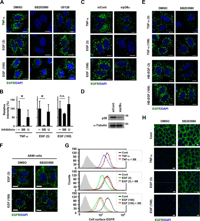
The canonical description of transmembrane receptor function is initial binding of ligand, followed by initiation of intracellular signaling and then internalization en route to degradation or recycling to the cell surface. It is known that low concentrations of extracellular ligand lead to a higher proportion of receptor that is recycled and that non-canonical mechanisms of receptor activation, including phosphorylation by the kinase p38, can induce internalization and recycling. However, no connections have been made between these pathways; i.e. it has yet to be established what happens to unbound receptors following stimulation with ligand. Here we demonstrate that a minimal level of activation of epidermal growth factor receptor (EGFR) tyrosine kinase by low levels of ligand is sufficient to fully activate downstream mitogen-activated protein kinase (MAPK) pathways, with most of the remaining unbound EGFR molecules being efficiently phosphorylated at intracellular serine/threonine residues by activated mitogen-activated protein kinase. This non-canonical, p38-mediated phosphorylation of the C-tail of EGFR, near Ser-1015, induces the clathrin-mediated endocytosis of the unliganded EGFR monomers, which occurs slightly later than the canonical endocytosis of ligand-bound EGFR dimers via tyrosine autophosphorylation. EGFR endocytosed via the non-canonical pathway is largely recycled back to the plasma membrane as functional receptors, whereas p38-independent populations are mainly sorted for lysosomal degradation. Moreover, ligand concentrations balance these endocytic trafficking pathways. These results demonstrate that ligand-activated EGFR signaling controls unliganded receptors through feedback phosphorylation, identifying a dual-mode regulation of the endocytic trafficking dynamics of EGFR.

Keywords: TNF-α; clathrin; endocytosis; epidermal growth factor receptor (EGFR); p38 MAPK; tumor necrosis factor.


Conflict of interest statement

The authors declare that they have no conflicts of interest with the contents of this article

Figures

Figure 1.
Figure 1.
p38-mediated endocytosis of EGFR with a ligand stimulation. A, HeLa cells were pretreated with 10 μm SB203580 or 5 μm U0126 for 30 min and then stimulated with 100 ng/ml TNF-α or 3 or 100 ng/ml EGF for another 15 min. The subcellular localization of EGFR was analyzed by confocal fluorescent microscopy. DAPI, 4′,6-diamidino-2-phenylindole. Scale bar =10 μm. B, the signal intensities of the internalized EGFR dots in A were calculated as a gray value. At least 50 cell profiles were counted, and data represent the mean ± S.D. *, p < 0.01, n.s., not significant. C and D, HeLa cells were transfected with siRNAs against p38α or the negative control and incubated for 48 h. Cells were stimulated with 20 ng/ml TNF-α or 3 or 100 ng/ml EGF for 15 min, and the subcellular localization of EGFR (green) was then analyzed by confocal fluorescent microscopy (C). Scale bars = 10 μm. The knockdown efficiency of p38 was assessed by immunoblotting (D). E, HeLa cells were pretreated with 10 μm SB203580 for 30 min and then stimulated with 3 or 100 ng/ml of TGF-α or HB-EGF for another 15 min. The subcellular localization of EGFR was analyzed. Scale bar =10 μm. F, A549 cells were pretreated with 10 μm SB203580 for 30 min and then stimulated with 3 or 100 ng/ml EGF for another 15 min. The subcellular localization of EGFR was analyzed. G and H, HeLa cells were pretreated with 10 μm SB203580 or 5 μm U0126 for 30 min and then stimulated with 100 ng/ml TNF-α or 3 or 100 ng/ml EGF for another 15 min. Scale bar =10 μm. Cell-surface EGFR expression was investigated by flow cytometry (G) and immunofluorescence (H). Cont, control. Scale bar =10 μm.
Figure 2.
Figure 2.
Ligand-induced feedback phosphorylation of EGFR by MAPKs. A, HeLa cells were pretreated with 1 μm PD153035 (PD), 10 μm SB, and 5 μm U for 30 min and then stimulated with 20 ng/ml TNF-α for another 10 min. Whole-cell lysates were separated by Zn2+ Phos tag SDS-PAGE, followed by immunoblotting (IB) with an anti-EGFR antibody. B and C, whole-cell lysates were prepared from HeLa cells stimulated with the indicated concentration of EGF for 10 min. After Zn2+ Phos tag SDS-PAGE (B) and normal SDS-PAGE (C), the expression of each protein was detected. D, the band densities in B and C were quantified by ImageJ software, and the band shift rate of EGFR in the Phos tag gel and Tyr(P)-1068–EGFR was calculated. Values represent the mean ± S.D. of three independent experiments as -fold increases. E, HeLa cells were stimulated with the indicated concentration of 3 ng/ml EGF for 10 min. Chemical inhibitors were pretreated for 30 min before addition of EGF. Whole-cell lysates were separated by Zn2+ Phos tag SDS-PAGE, followed by immunoblotting with an anti-EGFR antibody.
Figure 3.
Figure 3.
p38-mediated endocytosis of unliganded EGFR. A, HeLa cells were treated with the indicated concentration of rhodamine-EGF (Rh-EGF, red) for 15 min, fixed, and then stained with an anti-EGFR antibody (green). The subcellular localization of rhodamine and EGFR was examined by confocal fluorescent microscopy. Scale bars = 10 μm. DAPI, 4′,6-diamidino-2-phenylindole. B, the signal intensities of EGFR and rhodamine-EGF in (A) were calculated independently as gray values. At least 110 cell profiles were counted, and values represent the mean ± S.D. *, p < 0.01, n.s., not significant. C and D, HeLa cells were pretreated with 1 μm PD or 10 μm SB for 30 min and then treated with 3 or 100 ng/ml rhodamine-EGF (red) for another 15 min. The subcellular localization of EGFR (green) was assessed. Merged photos are shown. Scale bar =10 μm. E, the signal intensities in D were calculated. At least 110 cell profiles were counted, and values represent the mean ± S.D. *, p < 0.01.
Figure 4.
Figure 4.
Endocytosis of EGFR monomers via EGF-induced p38 activation. A, schematic of ligand-induced EGFR endocytosis. Ligand (red circle) binding to WT EGFR caused tyrosine phosphorylation (pY) of the EGFR asymmetric dimer. Activated WT EGFR induced the p38-dependent serine/threonine phosphorylation (pST) of dd-EGFR harboring a deletion (ΔCR1) and point mutations (I682Q and V924R). B and C, HeLa cells were transiently transfected with GFP-tagged dd-EGFR. Cells were stimulated with the indicated concentrations of EGF for 10 min (B). Transfected cells were pretreated with 10 μm SB203580 for 30 min and then treated with 3 or 100 ng/ml EGF for another 10 min (C). The Phos tag shift was assessed by immunoblotting (IB) with an anti-GFP antibody. D, HeLa cells were transiently transfected with GFP-tagged WT or dd-EGFR and stimulated with 100 ng/ml EGF for 10 min. The phosphorylation and total expression of endogenous (endo) and exogenous (GFP) EGFR were assessed by immunoblotting. E, CHO-K1 cells were transiently transfected with GFP-tagged WT or dd-EGFR and then stimulated with 100 ng/ml EGF for 15 min or 50 μm anisomycin for 30 min. The subcellular localization of EGFR-GFP was analyzed. Cont, control; DAPI, 4′,6-diamidino-2-phenylindole. Scale bar =10 μm. F and G, HeLa cells were transfected with GFP-tagged WT or dd-EGFR, pretreated with 10 μm SB203580 for 30 min, and then stimulated with 20 ng/ml TNF-α (F) or 5 ng/ml EGF (G). The subcellular localization of EGFR-GFP was analyzed. H and I, HeLa cells were transfected with siRNAs against CHC or the negative control. After a 48-h incubation, cells were further transfected with GFP-tagged WT or dd-EGFR and stimulated with 5 ng/ml EGF. Scale bar =10 μm. The knockdown efficiency of CHC was confirmed by immunoblotting (H). The subcellular localization of EGFR-GFP was analyzed (I). Scale bar =10 μm.
Figure 5.
Figure 5.
Phosphorylation of Ser/Thr is essential for non-canonical EGFR endocytosis. A, the structure and key amino acid residues of EGFR. Tyrosine phosphorylation sites and two p38 target regions are shown in red and green, respectively. TM, transmembrane domain; JM, juxtamembrane domain; TK, tyrosine kinase domain; CT, C-terminal domain. B, HeLa cells were transiently transfected with GFP-tagged dd-EGFR with the K721A mutation (dd-K721A), pretreated with 10 μm SB203580 for 30 min, and then stimulated with 3 ng ml−1 EGF or 20 ng/ml TNF-α for 15 min. The subcellular localization of EGFR-GFP was analyzed. Cont, control; DAPI, 4′,6-diamidino-2-phenylindole. Scale bar =10 μm. C, HeLa cells were transiently transfected with GFP-tagged dd-EGFR or dd-EGFR with the S1015/T1017/S1018A mutations in region 1 (R1m) or with the S1046/7A mutations in region 2 (R2m) and then stimulated with 5 ng/ml EGF for 15 min. The Phos tag shift was assessed by immunoblotting (IB) with an anti-GFP antibody. D, HeLa cells were transiently transfected with GFP-tagged dd-EGFR, dd+R1m, or dd+R2m and stimulated with 3 ng/ml EGF for 15 min. The subcellular localization of EGFR-GFP was analyzed. Scale bar =10 μm. E, CHO-K1 cells were transiently transfected with GFP-tagged WT EGFR or EGFR-R1m and stimulated with 100 ng/ml EGF for 15 min or anisomycin for 30 min. The subcellular localization of EGFR-GFP was analyzed. Scale bars = 10 μm.
Figure 6.
Figure 6.
Phosphorylation of EGFR at Ser-1015 in endocytic trafficking. A and C, HeLa cells were stimulated with 20 ng/ml TNF-α (A) or 3 ng/ml EGF (C) for the indicated time. B and D, HeLa cells were pretreated with 10 μm SB and 5 μm U for 30 min and then stimulated with 20 ng/ml TNF-α or 3 ng/ml EGF for another 10 min. A–D, whole-cell lysates were analyzed by immunoblotting with the indicated antibodies. E and F, HeLa cells were stimulated with 20 ng/ml TNF-α for the indicated time. The subcellular localization of total EGFR, Ser(P)-1015–EGFR, and Ser(P)-1047–EGFR was analyzed by confocal fluorescent microscopy. DAPI, 4′,6-diamidino-2-phenylindole. Scale bar =10 μm. G, HeLa cells were transiently transfected with GFP-tagged dd-EGFR and then stimulated with 3 ng/ml EGF or 20 ng/ml TNF-α for 15 min. The subcellular localization of EGFR-GFP and Ser(P)-1015-EGFR was analyzed. Scale bars = 10 μm. H, HeLa cells were transiently transfected with GFP-tagged WT-EGFR or dd-EGFR and stimulated with 5 ng/ml EGF for 5 or 15 min. The subcellular localization of EGFR-GFP was analyzed by confocal fluorescent microscopy. I, HeLa cells were stimulated with 20 ng/mL TNF-α or 3 ng/ml EGF for indicated time. The subcellular localization of total EGFR or Ser(P)-1015-EGFR was analyzed.
Figure 7.
Figure 7.
Post-endocytic fate of EGFR with low- and high-ligand stimuli. A, HeLa cells were treated with EGF (3 or 100 ng/ml) or TNF-α (20 ng/ml) for 15 or 60 min. The cell-surface expression of EGFR was analyzed by immunofluorescence under non-permeable conditions. Cont, control; DAPI, 4′,6-diamidino-2-phenylindole. Scale bar =10 μm. B, HeLa cells were pretreated with 3 ng/ml EGF at 37 °C for the indicated time, washed three times with cold PBS, and then treated with rhodamine-EGF (Rh-EGF) at 4 °C for 30 min. The cell-surface binding of rhodamine-EGF was analyzed by confocal fluorescence microscopy. Scale bar =10 μm. C, HeLa cells were treated with EGF or TGF-α (3, 10, 30, and 100 ng/ml) for 15 or 60 min. The cell-surface expression of EGFR was analyzed by flow cytometry, and the recycling ratio was calculated using the median values of fluorescence. Data are shown as the mean ± S.D. of three independent experiments. D and E, HeLa cells were stimulated with 10 ng/ml EGF or TGF-α for the indicated time in the absence or presence of 10 μm SB203580. The percentage of maximal internalization was calculated using the median values of fluorescence in flow cytometric assays. Data represent the mean ± S.D. of four (D) and three (E) independent experiments. *, p < 0.01. F and G, HeLa cells were transiently transfected with GFP-tagged WT EGFR or dd-EGFR and then stimulated with 100 ng/ml EGF for the indicated time. GFP-tagged and endogenous EGFR were detected by immunoblotting (IB) with an anti-EGFR antibody (F). The band densities of GFP-tagged and endogenous EGFR in control and stimulated (180 min) cells were measured (G). Data represent the mean ± S.D. of three independent experiments. *, p < 0.01. H, HeLa cells were transiently transfected with GFP-tagged WT or dd-EGFR and then stimulated with 100 ng/ml EGF for 15 and 180 min. The subcellular localization of EGFR-GFP was analyzed. Scale bars = 10 μm.
Figure 8.
Figure 8.
Transient suppression of ligand-induced EGFR activation. A, the protocol for the experiment in B is shown. B, HeLa cells were pretreated with 3 ng/ml EGF (pre-EGF) or 100 ng/ml TNF-α (pre-TNF-α) for 10 min and then stimulated with 100 ng/ml EGF for 2 or 5 min (post-EGF). C, HeLa cells were pretreated with 3 ng/ml EGF or 10 ng/ml TGF-α for the indicated time and then stimulated with 100 ng/ml EGF (post-EGF). Whole-cell lysates were immunoblotted with phospho-EGFR (Tyr-845, Tyr-1045, Ser-1046/7, and Tyr-1068), EGFR, and α-tubulin antibodies.
Figure 9.
Figure 9.
A model for ligand concentration–dependent dual endocytic trafficking of EGFR. Ligand binding induces dimerization of cell-surface EGFR and tyrosine phosphorylation-dependent endocytosis (canonical endocytosis, shown in red). In addition, canonically activated EGFR induces p38 activation, which leads to serine/threonine phosphorylation– and clathrin-dependent endocytosis of monomeric EGFR (non-canonical pathway, shown in green). Thus, ligand-induced EGFR trafficking involves the complex parallel events of canonical and non-canonical endocytosis, which are balanced by the ligand concentration, reflecting the initial ligand occupation rate. Under low-ligand conditions, a large amount of ligand-unbound EGFR is internalized via the non-canonical pathway and then recycled back to the cell surface. Conversely, ligand-occupied EGFR is internalized mainly via the canonical pathway under high-ligand conditions, which is preferentially sorted to the lysosome for degradation. CME, clathrin-mediated endocytosis; CIE, clathrin-independent endocytosis.

Similar articles

Cited by

References

    1. Avraham R., and Yarden Y. (2011) Feedback regulation of EGFR signaling: decision making by early and delayed loops. Nat. Rev. Mol. Cell Biol. 12, 104–117 10.1038/nrm3048 - DOI - PubMed
    1. Casaletto J. B., and McClatchey A. I. (2012) Spatial regulation of receptor tyrosine kinases in development and cancer. Nat. Rev. Cancer 12, 387–400 10.1038/nrc3277 - DOI - PMC - PubMed
    1. Yarden Y., and Pines G. (2012) The ERBB network: at last, cancer therapy meets systems biology. Nat. Rev. Cancer 12, 553–563 10.1038/nrc3309 - DOI - PubMed
    1. Parachoniak C. A., and Park M. (2012) Dynamics of receptor trafficking in tumorigenicity. Trends Cell Biol. 22, 231–240 10.1016/j.tcb.2012.02.002 - DOI - PubMed
    1. Goh L. K., and Sorkin A. (2013) Endocytosis of receptor tyrosine kinases. Cold. Spring. Harb. Perspect. Biol. 5, a017459 - PMC - PubMed

Publication types