. 2018 Feb;26(2):287-292.
doi: 10.1038/s41431-017-0007-0. Epub 2017 Dec 18.

CHARGE syndrome: a recurrent hotspot of mutations in CHD7 IVS25 analyzed by bioinformatic tools and minigene assays

Affiliations

CHARGE syndrome: a recurrent hotspot of mutations in CHD7 IVS25 analyzed by bioinformatic tools and minigene assays

Marine Legendre et al. Eur J Hum Genet. 2018 Feb.

Abstract

CHARGE syndrome is a rare genetic disorder mainly due to de novo and private truncating mutations of CHD7 gene. Here we report an intriguing hot spot of intronic mutations (c.5405-7G > A, c.5405-13G > A, c.5405-17G > A and c.5405-18C > A) located in CHD7 IVS25. Combining computational in silico analysis, experimental branch-point determination and in vitro minigene assays, our study explains this mutation hot spot by a particular genomic context, including the weakness of the IVS25 natural acceptor-site and an unconventional lariat sequence localized outside the common 40 bp upstream the acceptor splice site. For each of the mutations reported here, bioinformatic tools indicated a newly created 3' splice site, of which the existence was confirmed using pSpliceExpress, an easy-to-use and reliable splicing reporter tool. Our study emphasizes the idea that combining these two complementary approaches could increase the efficiency of routine molecular diagnosis.


Conflict of interest statement

The authors declare no conflict of interest

Figures
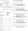
Fig. 1
Fig. 1
CHD7 IVS25 recurrent mutation analysis by minigene assays. A- Schematic representation of CHD7 exon 26 and its flanking regions. The last 21 nucleotides of IVS25 DNA sequence are indicated in lower cases and the consensus acceptor splice site in bold. The first nucleotides of exon 26 appear in upper case. Double arrows indicate different lengths in base pair. Simple arrows show the localization of PCR primers used for minigene generation (B1 and B2 refer to the attB1 and attB2 DNA sequences which were added in 5’ end of forward and reverse primers respectively). Overview of minigene assays using pSplice Express. Step 1 Amplification of the region of interest (amplicon of 624 bp). Two primers forward and reverse are used to amplify a part of the genomic DNA that harbors exon of interest (hatched rectangle) and its flanking intronic region (around 200 bp). The primers have recombination sites (AttB1, AttB2) that are indicated by circles. Step 2: Construction of the splicing reporter using pSpliceExpress. The PCR fragment is recombined in vitro with pSpliceExpress vector. The vector contains Cm and ccdB selection markers that are used to isolate recombined clones. The inserted DNA is flanked by two constitutive rat insulin exons, indicated by checkered rectangle. The transcript is driven by a RSV LTR promoter (full black arrow) and the subcloned genomic fragment flanked by attL sites, which are generated by the recombination of attB and attP sites. Step 3: Analysis of the splicing reporter. The minigene construct is transfected into HeLa cells. The RNA generated is determined by RT-PCR, using E2F and E3R primers (indicated by small arrows). The mRNA structures, indicated below the gene structure are expected to be generated by the construct. The range of amplicon length is indicated above double arrows. B- Minigene analysis of all recurrent IVS25 mutations by RT-PCR by fluorescent capillary electrophoresis. 380 to 407 bp PCR products correspond to CHD7 exon 26 minigenes, 261 bp amplicon corresponds to an alternative splicing between exon 2 and exon 3 of rat Insulin gene. C- cDNA organization and DNA sequencing of each RT-PCR amplicon obtained from each minigene analysis. Letters in bold indicate, for each minigene, the CHD7 protein sequence encoded by exon 26
Fig. 2
Fig. 2
CHD7 IVS25 branch point determination by lariat RT-PCR and minigene assays. A Nested lariat RT-PCR electrophoresis. Amplicons of 159 bp correspond to the relevant branch point sequence. The smaller amplicon that can be observed lane 3 (Fig. 2a) corresponds to an irrelevant product of splicing. B DNA sequencing of 159 bp amplicon obtained from nested lariat RT-PCR. A misincorporated “A” nucleotide (corresponding to the circled nucleotide T) indicates the branch point position. Arrows on the schematic explanation for lariat PCR indicate PCR primer localization: first round of lariat RT-PCR was performed with D and C, respectively located 163 nucleotide upstream CHD7-IVS25 3’ss and 97 bp upstream IVS2 rat Ins 5’ss. Nested PCR was performed using primers B and A, located respectively 125 bp upstream IVS25 3’ss and 38 bp downstream rats Ins 5’ss
Fig. 3
Fig. 3
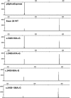
Selective disruption of branch point candidates using minigene assays. RT-PCR from each minigene is analyzed by fluorescent capillary electrophoresis. Lower amplicon (261 bp) corresponds to rat insulin minigene (exon 2 plus exon 3) and upper amplicon (391 bp) indicates full-length minigene containing the CHD7 sequence

References

    1. Janssen N, Bergman JEH, Swertz MA, et al. Mutation update on the CHD7 gene involved in CHARGE syndrome. Hum Mutat. 2012;33:1149–60. doi: 10.1002/humu.22086. - DOI - PubMed
    1. Kishore S, Khanna A, Stamm S. Rapid generation of splicing reporters with pSpliceExpress. Gene. 2008;427:104–10. doi: 10.1016/j.gene.2008.09.021. - DOI - PMC - PubMed
    1. Bilan F, Legendre M, Charraud V, et al. Complete screening of 50 patients with CHARGE syndrome for anomalies in the CHD7 gene using a denaturing high-performance liquid chromatography-based protocol: new guidelines and a proposal for routine diagnosis. J Mol Diagn JMD. 2012;14:46–55. doi: 10.1016/j.jmoldx.2011.08.003. - DOI - PubMed
    1. Sanlaville D, Etchevers HC, Gonzales M, et al. Phenotypic spectrum of CHARGE syndrome in fetuses with CHD7 truncating mutations correlates with expression during human development. J Med Genet. 2006;43:211–7. doi: 10.1136/jmg.2005.036160. - DOI - PMC - PubMed
    1. Gao K, Masuda A, Matsuura T, Ohno K. Human branch point consensus sequence is yUnAy. Nucleic Acids Res. 2008;36:2257–67. doi: 10.1093/nar/gkn073. - DOI - PMC - PubMed

Publication types

LinkOut - more resources