Review
. 2017 Dec 4:8:1708.
doi: 10.3389/fimmu.2017.01708. eCollection 2017.

The "Phagocytic Synapse" and Clearance of Apoptotic Cells

Affiliations
Review

The "Phagocytic Synapse" and Clearance of Apoptotic Cells

Nicole D Barth et al. Front Immunol. .

Abstract

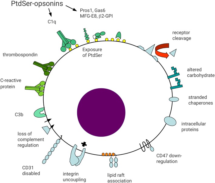
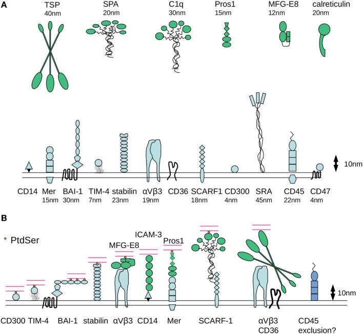
Apoptosis and subsequent phagocytic clearance of apoptotic cells is important for embryonic development, maintenance of tissues that require regular cellular renewal and innate immunity. The timely removal of apoptotic cells prevents progression to secondary necrosis and release of cellular contents, preventing cellular stress and inflammation. In addition, altered phagocyte behavior following apoptotic cell contact and phagocytosis engages an anti-inflammatory phenotype, which impacts upon development and progression of inflammatory and immune responses. Defective apoptotic cell clearance underlies the development of various inflammatory and autoimmune diseases. There is considerable functional redundancy in the receptors that mediate apoptotic cell clearance, highlighting the importance of this process in diverse physiological processes. A single phagocyte may utilize multiple receptor pathways for the efficient capture of apoptotic cells by phagocytes (tethering) and the subsequent initiation of signaling events necessary for internalization. In this review, we will consider the surface alterations and molecular opsonization events associated with apoptosis that may represent a tunable signal that confers distinct intracellular signaling events and hence specific phagocyte responses in a context-dependent manner. Efficient molecular communication between phagocytes and apoptotic targets may require cooperative receptor utilization and the establishment of efferocytic synapse, which acts to stabilize adhesive interactions and facilitate the organization of signaling platforms that are necessary for controlling phagocyte responses.

Keywords: apoptotic cells; cell–cell interactions; macrophage; opsonin; phagocytic receptor; phagocytosis; phosphatidylserine.


Figures

Figure 1
Figure 1
Schematic representation of surface molecular changes associated with apoptosis. Reduced cell surface molecule expression may occur through metalloprotease-mediated proteolytic shedding. Reduced receptor expression may act to limit apoptotic cell function and generate a distinct cell surface profile from viable cells. For example, reduced expression of CD47 or disabled CD31 may lead to loss of signals preventing phagocytosis. In addition, loss of integrin regulation may result in functional uncoupling in apoptotic cells, leading to cell detachment. Altered association of cell surface molecules with lipid rafts may alter functional activity, including gain-of-function of some receptors, e.g., FcγRIIa on myeloid cells. Altered carbohydrate processing may result in reduced sialic acid exposure and appearance of accessible mannose residues. Downregulation of complement regulatory molecules (e.g., CD55 and CD46) may lead to opsonization with complement components including C3b. Exposure of anionic phospholipids, including phosphatidylserine (PtdSer), allows binding of a broad range of opsonins to apoptotic cells. Protein S and Gas6 bind to PtdSer in a Ca2+-dependent manner, whereas milk fat globule EGF-factor 8 (MFG-E8) binds independent of Ca2+. Other less well defined apoptotic cell surface changes may allow binding of other opsonins including thrombospondin, C-reactive protein, and surfactant protein A. Finally, proteins with intracellular localizations may appear on the surface of apoptotic cells, including heat-shock proteins and calreticulin. Apoptotic cell surface molecules are shaded blue whereas apoptotic cell opsonins are shaded green.
Figure 2
Figure 2
Molecular dimensions of proteins involved in phagocytosis of apoptotic cells. (A) Some of the major families of receptors that have been identified as having a role in the phagocytosis of apoptotic cells are depicted, drawn approximately to scale based on available published crystallographic/NMR or cryo-electron microscopy data for various protein domains. One assumption made is that the molecules have a relatively rigid structure, although it is possible that flexibility would considerably alter axial length. There are considerable differences in the dimensions of apoptotic cell opsonins and in the receptors that mediate binding of phosphatidylserine (PtdSer) binding (either directly or indirectly). (B) Example receptor–counter-receptor pairings are shown to illustrate the likely differences in intermembrane “working” distance during phagocytosis, particularly for the molecules involved in the recognition of PtdSer (shown in yellow). Phagocyte receptors are shown in blue, opsonins are shown in green, and the apoptotic cell membrane is shown in red.

References

    1. Persson C, Uller L. Transepithelial exit of leucocytes: inflicting, reflecting or resolving airway inflammation? Thorax (2010) 65:1111–5.10.1136/thx.2009.133363 - DOI - PubMed
    1. Poon IKH, Lucas CD, Rossi AG, Ravichandran KS. Apoptotic cell clearance: basic biology and therapeutic potential. Nat Rev Immunol (2014) 14:166–80.10.1038/nri3607 - DOI - PMC - PubMed
    1. Lauber K, Blumenthal SG, Waibel M, Wesselborg S. Clearance of apoptotic cells: getting rid of the corpses. Mol Cell (2004) 14:277–87.10.1016/S1097-2765(04)00237-0 - DOI - PubMed
    1. Savill J, Dransfield I, Gregory C, Haslett C. A blast from the past: clearance of apoptotic cells regulates immune responses. Nat Rev Immunol (2002) 2:965–75.10.1038/nri957 - DOI - PubMed
    1. Elliott MR, Ravichandran KS. Clearance of apoptotic cells: implications in health and disease. J Cell Biol (2010) 189:1059–70.10.1083/jcb.201004096 - DOI - PMC - PubMed

LinkOut - more resources