. 2017 Oct;7(5):432-438.
doi: 10.21037/cdt.2017.06.10.

Coronary CT angiography-future directions

Affiliations

Coronary CT angiography-future directions

Stephan Achenbach. Cardiovasc Diagn Ther. 2017 Oct.

Abstract

Clinical applications of coronary CT angiography (CTA) will typically be based on the method´s very high sensitivity to identify coronary stenosis if image quality is good and if the pre-test likelihood of the patients is in the lower range. Guidelines of national and international cardiac societies are starting to incorporate coronary CTA into their recommendations for the management of patients with stable and acute chest pain. Initial data show that in the future, the use of coronary CTA may not only be able to replace other forms of diagnostic testing, but, in fact, may improve patient outcome. In this article, a perspective is provided on the future directions of coronary CTA.

Keywords: Coronary CT angiography (CTA); cardiac; flow; plaque.


Conflict of interest statement

Conflicts of Interest: The author has no conflicts of interest to declare.

Figures

Figure 1
Figure 1
High grade proximal stenosis of the left anterior descending coronary artery (arrows) in visualized in maximum intensity projection in transaxial orientation (A) and in a curved multiplanar reconstruction (B). Corresponding invasive angiogram (C).
Figure 2
Figure 2
Coronary CTA without stenoses, acquired at an effective radiation exposure of 0.7 mSv. Curved multiplanar reconstructions of all coronary arteries. CTA, CT angiography; LM, left main coronary artery; LAD, left anterior descending coronary artery; LCX, left circumflex coronary artery; RCA, right coronary artery.
Figure 3
Figure 3
Simulated FFR-CT based on fluid dynamic modeling. FFR-CT, fractional flow reserve determined through fluid dynamic modeling based on coronary CT angiography.
Figure 4
Figure 4
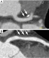
Visualization of non-obstructive plaque by coronary CTA. (A) Partly calcified plaque with positive remodeling in the left main coronary artery (arrows); (B) non-calcified plaque with positive remodeling in the proximal left anterior descending coronary artery. CTA, CT angiography.

References

    1. Moshage WE, Achenbach S, Seese B, et al. Coronary artery stenoses: three-dimensional imaging with electrocardiographically triggered, contrast agent-enhanced, electron-beam CT. Radiology 1995;196:707-14. 10.1148/radiology.196.3.7644633 - DOI - PubMed
    1. Schmermund A, Rensing BJ, Sheedy PF, et al. Intravenous electron-beam computed tomographic coronary angiography for segmental analysis of coronary artery stenoses. J Am Coll Cardiol 1998;31:1547-54. 10.1016/S0735-1097(98)00132-6 - DOI - PubMed
    1. Achenbach S, Moshage W, Ropers D, et al. Value of electron-beam computed tomography for the noninvasive detection of high-grade coronary-artery stenoses and occlusions. N Engl J Med 1998;339:1964-71. 10.1056/NEJM199812313392702 - DOI - PubMed
    1. Achenbach S, Ulzheimer S, Baum U, et al. Noninvasive coronary angiography by retrospectively ECG-gated multislice spiral CT. Circulation 2000;102:2823-8. 10.1161/01.CIR.102.23.2823 - DOI - PubMed
    1. Chinnaiyan KM, Boura JA, DePetris A, et al. Advanced Cardiovascular Imaging Consortium Coinvestigators Progressive radiation dose reduction from coronary computed tomography angiography in a statewide collaborative quality improvement program: results from the Advanced Cardiovascular Imaging Consortium. Circ Cardiovasc Imaging 2013;6:646-54. 10.1161/CIRCIMAGING.112.000237 - DOI - PubMed

LinkOut - more resources