

Advances in cardiac CT contrast injection and acquisition protocols
- PMID: 29255688
- PMCID: PMC5716940
- DOI: 10.21037/cdt.2017.06.07
Advances in cardiac CT contrast injection and acquisition protocols
Abstract
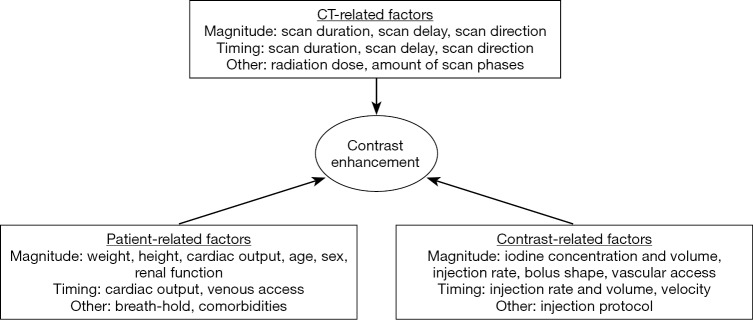
Cardiac computed tomography (CT) imaging has become an important part of modern cardiovascular care. Coronary CT angiography (CTA) is the first choice imaging modality for non-invasive visualization of coronary artery stenosis. In addition, cardiac CT does not only provide anatomical evaluation, but also functional and valvular assessment, and myocardial perfusion evaluation. In this article we outline the factors which influence contrast enhancement, give an overview of current contrast injection and acquisition protocols, with focus on current emerging topics such as pre-transcatheter aortic valve replacement (TAVR) planning, cardiac CT for congenital heart disease (CHD) patients, and myocardial CT perfusion (CTP). Further, we point out areas where we see potential for future improvements in cardiac CT imaging based on a closer interaction between CT scanner settings and contrast injection protocols to tailor injections to patient- and exam-specific factors.
Keywords: Contrast injection protocol; cardiac computed tomography (CT); coronary CT angiography (CTA); iodine contrast agent.
Conflict of interest statement
Conflicts of Interest: B Ghoshhajra is educational consultant for Siemens, Medtronic; JE Scholtz has no conflicts of interest to declare.
Figures
Similar articles
-
Practical considerations for optimizing cardiac computed tomography protocols for comprehensive acquisition prior to transcatheter aortic valve replacement.J Cardiovasc Comput Tomogr. 2016 Sep-Oct;10(5):364-74. doi: 10.1016/j.jcct.2016.07.007. Epub 2016 Jul 5. J Cardiovasc Comput Tomogr. 2016. PMID: 27475972 Review.
-
Coronary CT Angiography and Dual-Energy Computed Tomography in Ischemic Heart Disease Suspected Patients.Arch Iran Med. 2019 Jul 1;22(7):376-383. Arch Iran Med. 2019. PMID: 31679380
-
320-row CT transcatheter aortic valve replacement planning with a single reduced contrast media bolus injection.PLoS One. 2018 Sep 13;13(9):e0204145. doi: 10.1371/journal.pone.0204145. eCollection 2018. PLoS One. 2018. PMID: 30212567 Free PMC article.
-
Dynamic myocardial CT perfusion imaging-state of the art.Eur Radiol. 2023 Aug;33(8):5509-5525. doi: 10.1007/s00330-023-09550-y. Epub 2023 Mar 30. Eur Radiol. 2023. PMID: 36997751 Free PMC article. Review.
-
Low contrast medium-volume third-generation dual-source computed tomography angiography for transcatheter aortic valve replacement planning.Eur Radiol. 2017 May;27(5):1944-1953. doi: 10.1007/s00330-016-4537-6. Epub 2016 Aug 23. Eur Radiol. 2017. PMID: 27553939
Cited by
-
Cardiac CT for Measurement of Right Ventricular Volume and Function in Comparison with Cardiac MRI: A Meta-Analysis.Korean J Radiol. 2020 Apr;21(4):450-461. doi: 10.3348/kjr.2019.0499. Korean J Radiol. 2020. PMID: 32193893 Free PMC article.
-
Dynamic CT myocardial perfusion without image registration.Sci Rep. 2022 Jul 23;12(1):12608. doi: 10.1038/s41598-022-16573-w. Sci Rep. 2022. PMID: 35871187 Free PMC article.
-
The reproducibility and predictivity of radiomic features extracted from dynamic contrast-enhanced computed tomography of hepatocellular carcinoma.PLoS One. 2024 Sep 13;19(9):e0310486. doi: 10.1371/journal.pone.0310486. eCollection 2024. PLoS One. 2024. PMID: 39269960 Free PMC article.
-
Feasibility of Non-Invasive Coronary Artery Disease Screening with Coronary CT Angiography before Transcatheter Aortic Valve Implantation.J Clin Med. 2023 Mar 15;12(6):2285. doi: 10.3390/jcm12062285. J Clin Med. 2023. PMID: 36983286 Free PMC article.
-
Automatic 3D coronary artery segmentation based on local region active contour model.J Thorac Dis. 2024 Apr 30;16(4):2563-2579. doi: 10.21037/jtd-24-421. Epub 2024 Apr 28. J Thorac Dis. 2024. PMID: 38738249 Free PMC article.
References
-
- Hutchison SJ, Merchant N. Principles of Cardiac and Vascular Computed Tomography: Expert Consult. Elsevier Health Sciences, 2014.
LinkOut - more resources
Full Text Sources
Other Literature Sources