. 2017 Dec 13;3(12):eaao2267.
doi: 10.1126/sciadv.aao2267. eCollection 2017 Dec.

A mixed-kinetic model describes unloaded velocities of smooth, skeletal, and cardiac muscle myosin filaments in vitro

Affiliations

A mixed-kinetic model describes unloaded velocities of smooth, skeletal, and cardiac muscle myosin filaments in vitro

Richard K Brizendine et al. Sci Adv. .

Abstract

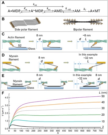
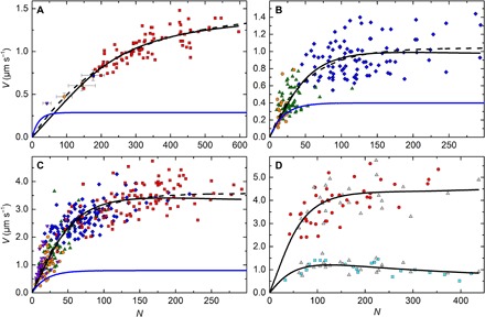
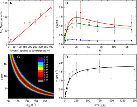
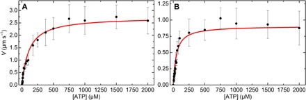
In vitro motility assays, where purified myosin and actin move relative to one another, are used to better understand the mechanochemistry of the actomyosin adenosine triphosphatase (ATPase) cycle. We examined the relationship between the relative velocity (V) of actin and myosin and the number of available myosin heads (N) or [ATP] for smooth (SMM), skeletal (SKM), and cardiac (CMM) muscle myosin filaments moving over actin as well as V from actin filaments moving over a bed of monomeric SKM. These data do not fit well to a widely accepted model that predicts that V is limited by myosin detachment from actin (d/ton), where d equals step size and ton equals time a myosin head remains attached to actin. To account for these data, we have developed a mixed-kinetic model where V is influenced by both attachment and detachment kinetics. The relative contributions at a given V vary with the probability that a head will remain attached to actin long enough to reach the end of its flexible S2 tether. Detachment kinetics are affected by L/ton, where L is related to the tether length. We show that L is relatively long for SMM, SKM, and CMM filaments (59 ± 3 nm, 22 ± 9 nm, and 22 ± 2 nm, respectively). In contrast, L is shorter (8 ± 3 nm) when myosin monomers are attached to a surface. This suggests that the behavior of the S2 domain may be an important mechanical feature of myosin filaments that influences unloaded shortening velocities of muscle.


Figures

Fig. 1
Fig. 1. Kinetic scheme, myosin structures, motility assay geometries, and model predictions.
(A) Kinetic scheme for myosin (M) attachment to actin (A). D, ADP; T, ATP; Pi, phosphate. Kw, equilibrium constant for weak binding of myosin to actin; kws, forward rate constant for the weak to strong transition. We assume that −kws is insignificant. See text for other rate constants. (B to E) None of the cartoons are to scale. (B) Side polar (smooth muscle) and bipolar myosin filaments (skeletal and cardiac muscle), heads (orange), and tails (black). (C) A/Mm assay with actin filament (green) being moved by myosin heads (orange) attached to coverslip (blue). Only the S2 region of the tail (black) is shown. Before the working step (left), after the working step (middle), and after the fullest possible extension of S2 (right) by other heads undergoing the working steps (not shown for clarity). In this example, d = L = 8 nm. (D) Mf/A assay (5, 6), myosin filament (orange) moves over biotinylated actin (green) attached to PEG brush-coated coverslip (not shown). Pre-working step (left), post-working step (middle), and after full extension of S2 (right) by other working heads (not shown) giving L = 32 nm in this example. This head is now a drag head and therefore must detach from actin before further movement of the filament is possible. See movies S1 and S2 and the study of Brizendine et al. (6) for a more complete description. (E) A/Mf assay, a portion of a bipolar biotinylated myosin filament (black) attached via streptavidin to a biotinylated PEG brush-coated coverslip with physiological or fast (left) and nonphysiological or slow head-actin interactions (right). The slow heads need to spin or swivel to attach to actin, represented by a loop in the S2 domain, but the mechanism is unknown. Table 2 shows that L for the slow heads is finite, but the structural basis is unknown and therefore not depicted. (F) Mixed-kinetic model predictions at varying L’s. Plots predicted by Eq. 5 for katt = 6 s−1, d = 8 nm, kAD = 100 s−1, kT = 2 μM−1 s−1, [ATP] = 1 mM. Please see Methods for an Excel spreadsheet link to generate additional curves.
Fig. 2
Fig. 2. Detachment-limited model (Eq. 1) and mixed-kinetic model (Eq. 5) fits to N dependence of filament velocities (V) at saturating [ATP].
All experiments were performed in the observation buffer at 30°C. (A) SMM filaments moving in the Mf/A assay at 0.75 to 2.0 mM ATP from Brizendine et al. (6). Purple triangles, 26% SMM co-filaments, n = 10; orange circles, 51% SMM co-filaments; green triangles, 75% SMM co-filaments; blue diamond, rapidly diluted (short) SMM filaments; each point is the average V and N from 10 individual filament trajectories, and error bars show the SD. Red squares, SMM filaments prepared by dialysis; each point (n = 82) is V of a single filament trajectory. (B) CMM filaments moving in the Mf/A assay at 0.75 to 2.0 mM ATP. Blue diamonds, CMM filaments, n = 95; green triangles, 50% CMM co-filaments, n = 62; orange circles, 30% CMM co-filaments, n = 19. (C) SKM filaments moving in the Mf/A assay at 0.75 to 2.0 mM ATP. Purple triangles, 25% SKM co-filaments, n = 38; orange circles, 50% SKM co-filaments, n = 47; green triangles, 75% SKM co-filaments, n = 41; blue diamonds, SKM filaments prepared by rapid dilution (short), n = 122; red squares, SKM filaments prepared by dialysis. Each point (n = 102) is V of a single filament trajectory. For (A) to (C): Fits to Eq. 1 with d fixed at 8 nm (dashed lines) and d = 8 nm, r = 0.05, and kAD = 36, 50, and 100 s−1, respectively, for (A), (B), and (C) (blue). See Table 1 for a summary of fit parameters. (D) V of actin moving on SKM filaments in the A/Mf assay at 1 mM ATP. Gray triangles, fast and slow V measured from a single actin filament trajectory moving across a single myosin filament (n = 25); red circles, V from a single actin filament trajectory (n = 42); cyan squares, V from a single actin filament trajectory (n = 21). All plots show a fit to Eq. 5 (black lines). See Table 2 for a summary of fit parameters.
Fig. 3
Fig. 3. A/Mm data for SKM.
All experiments were performed in the observation buffer at 30°C. (A) Relationship between [myosin] applied to the coverslip (μg ml−1) and N. Red points are averages of two to six independent experiments; error bars show the SD; the red line is a linear fit, slope = 0.34 N ml μg−1. Black squares, data from Harris and Warshaw (17). (B) N dependence of V at 1 mM (red), 100 μM (green), and 20 μM ATP (blue). N values correspond to applied SKM concentrations between 5 and 400 μg ml−1. (C) Contour plot of L versus kAD showing dependence of R2 values from the fit to 1000 μM ATP data from (B) (see fig. S2 for plots of 20 and 100 μM ATP data). Color scale shows the range of R2 values. (D) ATP dependence of V at N = 34 (100 μg ml−1). For (B) and (D), each point is an average of 40 individual actin filament trajectories, and error bars show the SD. Respective lines show the best fits to Eq. 5.
Fig. 4
Fig. 4. ATP dependence of filament V at fixed N from the Mf/A assay.
(A) SKM filaments formed by rapid dilution. (B) CMM filaments formed by rapid dilution. For both plots, each point is an average of 40 individual trajectories. Error bars show the SD. Red lines show the fits to Eq. 5 (see Table 2 for results). R2 = 0.95 and 0.96, respectively. All experiments were performed in the observation buffer at 30°C.

References

    1. Kron S. J., Spudich J. A., Fluorescent actin filaments move on myosin fixed to a glass surface. Proc. Natl. Acad. Sci. U.S.A. 83, 6272–6276 (1986). - PMC - PubMed
    1. Yanagida T., Nakase M., Nishiyama K., Oosawa F., Direct observation of motion of single F-actin filaments in the presence of myosin. Nature 307, 58–60 (1984). - PubMed
    1. Toyoshima Y. Y., Kron S. J., McNally E. M., Niebling K. R., Toyoshima C., Spudich J. A., Myosin subfragment-1 is sufficient to move actin filaments in vitro. Nature 328, 536–539 (1987). - PubMed
    1. Yengo C. M., Takagi Y., Sellers J. R., Temperature dependent measurements reveal similarities between muscle and non-muscle myosin motility. J. Muscle Res. Cell Motil. 33, 385–394 (2012). - PMC - PubMed
    1. Haldeman B. D., Brizendine R. K., Facemyer K. C., Baker J. E., Cremo C. R., The kinetics underlying the velocity of smooth muscle myosin filament sliding on actin filaments in vitro. J. Biol. Chem. 289, 21055–21070 (2014). - PMC - PubMed

Publication types

LinkOut - more resources