Review
. 2018 Apr;410(10):2467-2484.
doi: 10.1007/s00216-017-0772-1. Epub 2017 Dec 18.

Recent mass spectrometry-based techniques and considerations for disulfide bond characterization in proteins

Affiliations
Review

Recent mass spectrometry-based techniques and considerations for disulfide bond characterization in proteins

Jude C Lakbub et al. Anal Bioanal Chem. 2018 Apr.

Abstract

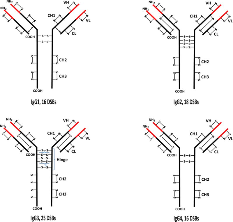
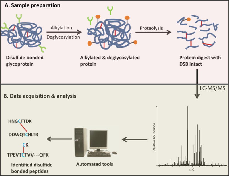
Disulfide bonds are important structural moieties of proteins: they ensure proper folding, provide stability, and ensure proper function. With the increasing use of proteins for biotherapeutics, particularly monoclonal antibodies, which are highly disulfide bonded, it is now important to confirm the correct disulfide bond connectivity and to verify the presence, or absence, of disulfide bond variants in the protein therapeutics. These studies help to ensure safety and efficacy. Hence, disulfide bonds are among the critical quality attributes of proteins that have to be monitored closely during the development of biotherapeutics. However, disulfide bond analysis is challenging because of the complexity of the biomolecules. Mass spectrometry (MS) has been the go-to analytical tool for the characterization of such complex biomolecules, and several methods have been reported to meet the challenging task of mapping disulfide bonds in proteins. In this review, we describe the relevant, recent MS-based techniques and provide important considerations needed for efficient disulfide bond analysis in proteins. The review focuses on methods for proper sample preparation, fragmentation techniques for disulfide bond analysis, recent disulfide bond mapping methods based on the fragmentation techniques, and automated algorithms designed for rapid analysis of disulfide bonds from liquid chromatography-MS/MS data. Researchers involved in method development for protein characterization can use the information herein to facilitate development of new MS-based methods for protein disulfide bond analysis. In addition, individuals characterizing biotherapeutics, especially by disulfide bond mapping in antibodies, can use this review to choose the best strategies for disulfide bond assignment of their biologic products. Graphical Abstract This review, describing characterization methods for disulfide bonds in proteins, focuses on three critical components: sample preparation, mass spectrometry data, and software tools.

Keywords: Disulfide bond; Liquid chromatography; Mass spectrometry; Software.


Conflict of interest statement

The authors of the paper declare that they have no conflicts of interest.

Figures

Figure 1
Figure 1
Structures showing the typical disulfide bond (DSB) patterns of the four classes of immunoglobulin gamma (IgG) antibodies. The black and red parts represent the constant (C) and variable (V) regions, respectively. H represents the heavy chain while L represents the light chain. VL and CL are domains on the light chain while VH, CH1, hinge, CH2, and CH3 are domains on the heavy chain. IgG3 has a 15-residue segment in the hinge region that is repeated 3 times; each of the segments contains 3 disulfide bonds.
Figure 2
Figure 2
Disulfide bond analysis workflow. A. Sample preparation of non-reduced protein digest for disulfide bond analysis. Alkylation of free Cys is important to prevent disulfide shuffling (see text, Section 2.3). For glycoproteins, deglycosylation would ensure less complex data (see text, Section 2.4). B. Disulfide bond assignment from LC-MS/MS data.
Figure 3
Figure 3
Schematic illustration of the different possible routes for formation of non-native disulfide bonds during sample preparation. Purple Cys residues are shown in native disulfide conformation; Cys residues marked with asterisks are modified into non-native conformations (red Cys residues). A. Reaction between two free Cys residues. B. Reaction between free Cys residues and Cys residues involved in a disulfide bond. C. Reaction between Cys residues that were formerly involved in disulfide bonds.
Figure 4
Figure 4
CID (A) and ETD (B) spectra of a disulfide bonded peptide from the CH2 domain of IgG3 monoclonal antibody. The spectra show the characteristic b/y (A) and c/z (B) ions resulting from the backbone cleavage of the peptides linked by the disulfide bond (DSB). Fragment ions not containing the DSB are labeled in green, and those containing the intact DSB are labeled in red. CID fragment ions resulting from two cleavage events and containing the intact DSB are in brackets. In the ETD spectrum (B), intense peaks of the Cys-containing peptides linked by the disulfide bond are observed at m/z 250.4 and 1179.3. These peaks result from the cleavage of the DSB by ETD, and they are not observed in (A).
Figure 5
Figure 5
Schematic representation of bottom-up approaches for disulfide bond analysis. The methods generally fall under two broad categories: (1) disulfide bond mapping from protein digests with reduced and non-reduced disulfide bonds (profile comparison), and (2) disulfide bond mapping from only intact (non-reduced) protein digests. Non-reduced analysis can be further divided into methods that involve post-column reduction of disulfide bonds, reduction of the disulfide bonds in the gas phase (ETD and EThcD fragmentation methods), and those that do not require reduction of the disulfide bonds (CID and HCD fragmentation methods). Data analysis after post-column reduction has not been automated yet.
Figure 6
Figure 6
Example of Extracted Ion Chromatograms (XICs) used to identify disulfide linkages in an IgG antibody. Correct disulfide linkages are highlighted in blue. Aberrant linkages are marked with asterisks. Reproduced from Ref. with permission from The Royal Society of Chemistry.
Figure 7
Figure 7
Rapid assignment of disulfide linkage via a1 ion recognition (RADAR), a1 ions are used to identify potential disulfides which are then confirmed using intact molecular weight and b/y ions. A) Experimental workflow for RADAR analysis. B) MS2 spectra used to identify disulfide bonded peptides. Adapted with permission from Ref. . Copyright 2012 American Chemical Society.

References

    1. Woycechowsky KJ, Raines RT. Native Disulfide Bond Formation in Proteins. Curr Opin Chem Biol. 2000;4(5):533–539. - PMC - PubMed
    1. Wilkinson B, Gilbert HF. Protein Disulfide Isomerase. Biochim Biophys Acta. 2004;1699(1–2):35–44. - PubMed
    1. Butera D, Cook KM, Chiu J, Wong JW, Hogg PJ. Control of Blood Proteins by Functional Disulfide Bonds. Blood. 2014;123(13):2000–2007. - PMC - PubMed
    1. Farrah T, Deutsch EW, Omenn GS, Campbell DS, Sun Z, Bletz JA, Mallick P, Katz JE, Malmstrom J, Ossola R, Watts JD, Lin B, Zhang H, Moritz RL, Aebersold R. A High-Confidence Human Plasma Proteome Reference Set with Estimated Concentrations in PeptideAtlas. Mol Cell Proteomics. 2011;10(9):M110.006353. - PMC - PubMed
    1. Wypych J, Li M, Guo A, Zhang Z, Martinez T, Allen MJ, Fodor S, Kelner DN, Flynn GC, Liu YD, Bondarenko PV, Ricci MS, Dillon TM, Balland A. Structural and Functional Characterization of Disulfide Isoforms of the Human IgG2 Subclass. J Biol Chem. 2008;283(23):16194–16205. - PMC - PubMed