

Lipid environment of membrane proteins in cryo-EM based structural analysis
- PMID: 29256118
- PMCID: PMC5899730
- DOI: 10.1007/s12551-017-0371-6
Lipid environment of membrane proteins in cryo-EM based structural analysis
Abstract
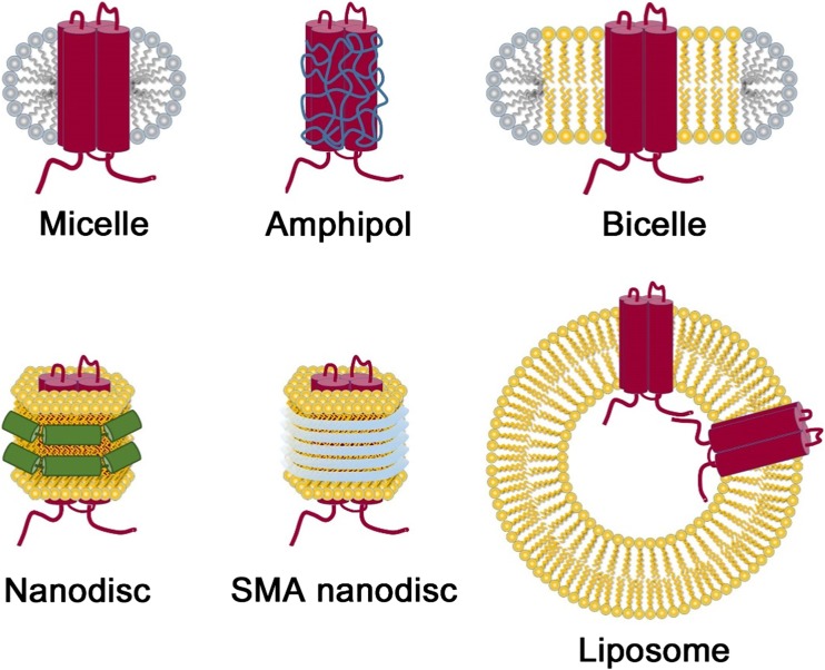
Cryoelectron microscopy (cryo-EM) in association with a single particle analysis method (SPA) is now a promising tool to determine the structures of proteins and their macromolecular complexes. The development of direct electron detection cameras and image processing technologies has allowed the structures of many important proteins to be solved at near-atomic resolution or, in some cases, at atomic resolution, by overcoming difficulties in crystallization or low yield of protein production. In the case of membrane-integrated proteins, the proteins were traditionally solubilized and stabilized with various kind of detergents. However, the density of detergent micelles diminished the contrast of membrane proteins in cryo-EM studies and made it difficult to obtain high-resolution structures. To improve the resolution of membrane protein structures in cryo-EM studies, major improvements have been made both in sample preparation techniques and in hardware and software developments. The focus of our review is on improvements which have been made in the various techniques for sample preparation for cryo-EM studies, with a specific interest placed on techniques for mimicking the lipid environment of membrane proteins.
Keywords: Cryoelectron microscopy; Lipid environment; Membrane proteins; Single particle analysis.
Conflict of interest statement
Conflict of interest
Kazuhiro Mio declares that he has no conflicts of interest. Chikara Sato declares that he has no conflicts of interest.
Ethical approval
This article does not contain any studies with human participants or animals performed by any of the authors.
Figures


Similar articles
-
Sample preparation of biological macromolecular assemblies for the determination of high-resolution structures by cryo-electron microscopy.Microscopy (Oxf). 2016 Feb;65(1):23-34. doi: 10.1093/jmicro/dfv367. Epub 2015 Dec 15. Microscopy (Oxf). 2016. PMID: 26671943 Review.
-
Retrospect and Prospect of Single Particle Cryo-Electron Microscopy: The Class of Integral Membrane Proteins as an Example.J Chem Inf Model. 2020 May 26;60(5):2448-2457. doi: 10.1021/acs.jcim.9b01015. Epub 2020 Mar 20. J Chem Inf Model. 2020. PMID: 32163280 Review.
-
Near-Atomic Resolution Cryo-EM Image Reconstruction of RNA.Methods Mol Biol. 2023;2568:179-192. doi: 10.1007/978-1-0716-2687-0_12. Methods Mol Biol. 2023. PMID: 36227569
-
Structural Analysis of Protein Complexes by Cryo-Electron Microscopy.Methods Mol Biol. 2024;2715:431-470. doi: 10.1007/978-1-0716-3445-5_27. Methods Mol Biol. 2024. PMID: 37930544 Review.
-
Overview of Membrane Protein Sample Preparation for Single-Particle Cryo-Electron Microscopy Analysis.Int J Mol Sci. 2023 Sep 30;24(19):14785. doi: 10.3390/ijms241914785. Int J Mol Sci. 2023. PMID: 37834233 Free PMC article. Review.
Cited by
-
Foreword to 'Multiscale structural biology: biophysical principles and mechanisms underlying the action of bio-nanomachines', a special issue in Honour of Fumio Arisaka's 70th birthday.Biophys Rev. 2018 Apr;10(2):105-129. doi: 10.1007/s12551-018-0401-z. Epub 2018 Mar 2. Biophys Rev. 2018. PMID: 29500796 Free PMC article.
-
Comparing ATPase activity of ATP-binding cassette subfamily C member 4, lamprey CFTR, and human CFTR using an antimony-phosphomolybdate assay.Front Pharmacol. 2024 Feb 19;15:1363456. doi: 10.3389/fphar.2024.1363456. eCollection 2024. Front Pharmacol. 2024. PMID: 38440176 Free PMC article.
-
Importance of molecular dynamics equilibrium protocol on protein-lipid interaction near channel pore.Biophys Rep (N Y). 2022 Sep 29;2(4):100080. doi: 10.1016/j.bpr.2022.100080. eCollection 2022 Dec 14. Biophys Rep (N Y). 2022. PMID: 36425669 Free PMC article.
-
New Structures and Gating of Voltage-Dependent Potassium (Kv) Channels and Their Relatives: A Multi-Domain and Dynamic Question.Int J Mol Sci. 2019 Jan 10;20(2):248. doi: 10.3390/ijms20020248. Int J Mol Sci. 2019. PMID: 30634573 Free PMC article. Review.
-
Regulation of Membrane Calcium Transport Proteins by the Surrounding Lipid Environment.Biomolecules. 2019 Sep 20;9(10):513. doi: 10.3390/biom9100513. Biomolecules. 2019. PMID: 31547139 Free PMC article. Review.
References
-
- Adrian M, Dubochet J, Lepault J, McDowall AW. Cryo-electron microscopy of viruses. Nature. 1984;308:32–36. - PubMed
-
- Alpes H, Apell H-J, Knoll G, Plattner H, Riek R. Reconstitution of Na+/K+-ATPase into phosphatidylcholine vesicles by dialysis of nonionic alkyl maltoside detergents. Biochim Biophys Acta. 1988;946:379–388. - PubMed
-
- Arachea BT, Sun Z, Potente N, Malik R, Isailovic D, Viola RE. Detergent selection for enhanced extraction of membrane proteins. Protein Expr Purif. 2012;86:12–20. - PubMed
Publication types
LinkOut - more resources
Full Text Sources
Other Literature Sources
