. 2018 Apr;67(4):537-549.
doi: 10.1007/s00262-017-2106-8. Epub 2017 Dec 18.

IL-6 and IL-10 are associated with good prognosis in early stage invasive breast cancer patients

Affiliations

IL-6 and IL-10 are associated with good prognosis in early stage invasive breast cancer patients

Narmeen Ahmad et al. Cancer Immunol Immunother. 2018 Apr.

Abstract

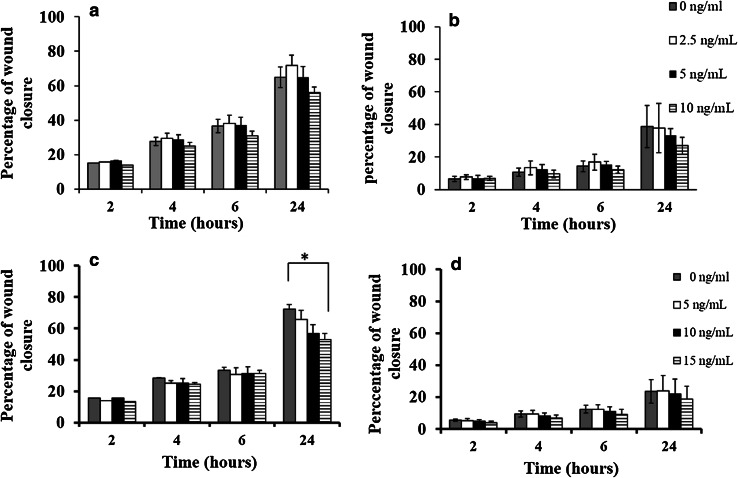
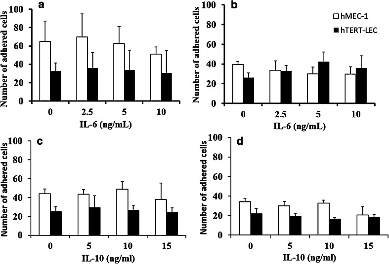
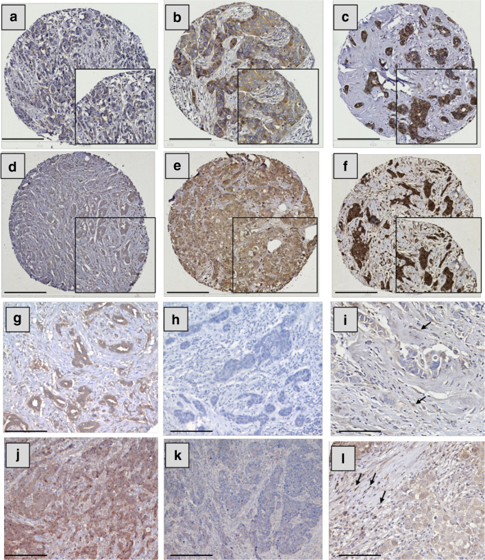
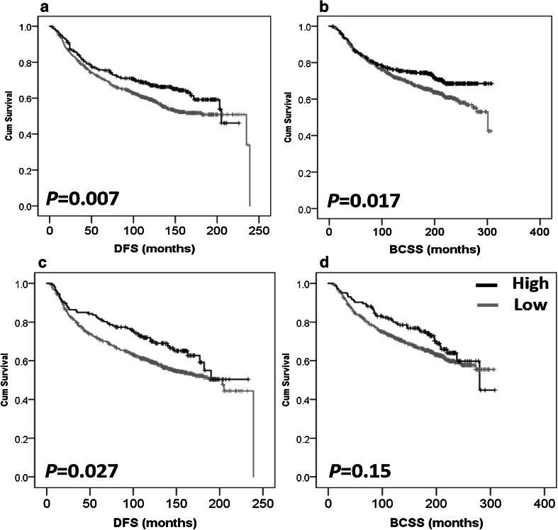
Macrophage-associated cytokines play an important role in cancer metastasis; however, the functions of interleukins (IL) 6 and 10 in breast cancer (BC) progression and metastasis are not clear. In this study the roles of IL-6/IL-10 in regulating vascular invasion and their prognostic significance in BC are investigated. MDA-MB-231 and MCF-7 migration (± IL-6 or IL-10) was assessed by scratch wound assay. Cancer cell adhesion to IL-6/IL-10 stimulated blood and lymphatic endothelial cells (EC) was investigated. Expression of IL-6 /IL-10 was assessed using immunohistochemistry in an annotated cohort of early stage BC (n = 1380) and associations with clinicopathological variables and clinical outcome evaluated. IL-6 did not alter BC cell migration however a dose-dependent inhibition in MDA-MB-231 migration with IL-10 treatment was observed (P = 0.03). BC cells were more adhesive to blood vs lymphatic EC, however, IL-6/IL-10 had no effect on adhesion patterns. High expression of IL-6/IL-10 was associated with clinicopathological criteria (e.g. hormone receptor status, all P < 0.05), improved disease-free survival (DFS; P < 0.05) and improved BC-specific survival (BCSS; only IL-6, P = 0.017). However, neither IL-6 nor IL-10 expression were independent prognostic factors from multivariate analysis. In BC subgroups, IL-6 and IL-10 were good prognosticators in terms of DFS in non-basal, non-triple-negative (non-TN), ER-positive, PgR-positive (only IL-10), and Her-2-negative (only IL-6) BC (all P < 0.05). IL-6 was associated with improved BCSS in non-basal, ER-positive and non-TN BC (all P < 0.05).

Keywords: Breast cancer; IL-10; IL-6; Macrophage-associated cytokines; Metastasis.


Conflict of interest statement

The authors declare that they have no competing interest.

Figures

Fig. 1
Fig. 1
Effect of recombinant human IL-6 or IL-10 stimulation on breast cancer cell migration rate. IL-6 treatment caused a slight decrease in MDA-MB-231 (a) and MCF-7 (b) migration after 24 h treatment; however, the reduction in migration did not reach statistical significance. IL-10 (15 ng/mL) was associated with a significant reduction in MDA-MB-231 migration (P = 0.03) (c), but has no significant effect on MCF-7 migration (d). Data are presented as mean ± SD of three independent experiments each carried out in triplicate, and P values evaluated by paired sample t test (asterisk represent a significant P value)
Fig. 2
Fig. 2
Effect of IL-6 and IL-10 on endothelial adhesion patterns of MDA-MB-231 and MCF-7. IL-6 or IL-10 stimulation of blood (hMEC-1) and lymphatic (hTERT-LEC) endothelial cells did not significantly alter adhesion patterns compared to the unstimulated controls. a MDA-MB-231 and b MCF-7 adhesion to IL-6 stimulated endothelial cells, c MDA-MB-231 and d MCF-7 adhesion to IL-10 stimulated endothelia. Data represent the mean of adhered cells ± SD of three independent experiments, each carried out in duplicate (n = 6)
Fig. 3
Fig. 3
IL-6 and IL-10 expression in breast cancer and stromal cells. Representative images of tumour staining with IL-6 (ac) and IL-10 (df). Staining pattern: a, d weak, b, e moderate, and c, f strong. g, h, j, k Representative images of specificity tests of IL-6 and IL-10 antibodies, respectively: BC staining with IL-6 antibody (g) or IL-6 antibody blocked overnight with 1 µg of rIL-6 (h); BC staining with IL-10 antibody alone (j) or IL-10 antibody blocked overnight with 2 µg of rIL-10 (k). Examples of stromal expression of IL-6 and IL-10 are shown in i, l, respectively (black arrows). Photomicrographs: af; × 100 and gl × 200 magnification; inset boxes at × 200 magnification; scale bars 100 µm
Fig. 4
Fig. 4
Kaplan–Meier analysis of association between IL-6 and IL-10 expression with breast cancer prognosis. High IL-6 expression is significantly associated with improved disease-free survival (P = 0.007, a) and improved breast cancer-specific survival (P = 0.017, b). High IL-10 expression is significantly associated with improved disease-free survival (P = 0.027, c) but not breast cancer-specific survival (P = 0.150, d). Significance was determined using the log-rank test. Black represents high expression and grey represents low expression of the cytokine

References

    1. Mohammed RA, Ellis IO, Elsheikh S, Paish EC, Martin SG. Lymphatic and angiogenic characteristics in breast cancer: morphometric analysis and prognostic implications. Breast Cancer Res Treat. 2009;113(2):261–273. doi: 10.1007/s10549-008-9936-1. - DOI - PubMed
    1. Mohammed RA, Menon S, Martin SG, Green AR, Paish EC, Ellis IO. Prognostic significance of lymphatic invasion in lymph node-positive breast carcinoma: findings from a large case series with long-term follow-up using immunohistochemical endothelial marker. Mod Pathol. 2014;27(12):1568–1577. doi: 10.1038/modpathol.2014.60. - DOI - PubMed
    1. Mohammed ZM, Going JJ, Edwards J, Elsberger B, Doughty JC, McMillan DC. The relationship between components of tumour inflammatory cell infiltrate and clinicopathological factors and survival in patients with primary operable invasive ductal breast cancer. Br J Cancer. 2012;107(5):864–873. doi: 10.1038/bjc.2012.347. - DOI - PMC - PubMed
    1. Pollard JW. Macrophages define the invasive microenvironment in breast cancer. J Leukoc Biol. 2008;84(3):623–630. doi: 10.1189/jlb.1107762. - DOI - PMC - PubMed
    1. Grivennikov SI, Greten FR, Karin M. Immunity, inflammation, and cancer. Cell. 2010;140(6):883–899. doi: 10.1016/j.cell.2010.01.025. - DOI - PMC - PubMed