

Functional Reorganization of Right Prefrontal Cortex Underlies Sustained Naming Improvements in Chronic Aphasia via Repetitive Transcranial Magnetic Stimulation
- PMID: 29256908
- PMCID: PMC5797702
- DOI: 10.1097/WNN.0000000000000141
Functional Reorganization of Right Prefrontal Cortex Underlies Sustained Naming Improvements in Chronic Aphasia via Repetitive Transcranial Magnetic Stimulation
Abstract
Background and objective: While noninvasive brain stimulation techniques show promise for language recovery after stroke, the underlying mechanisms remain unclear. We applied inhibitory repetitive transcranial magnetic stimulation (rTMS) to regions of interest in the right inferior frontal gyrus of patients with chronic poststroke aphasia and examined changes in picture naming performance and cortical activation.
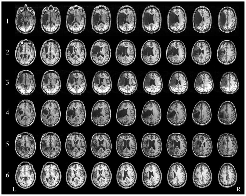
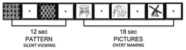
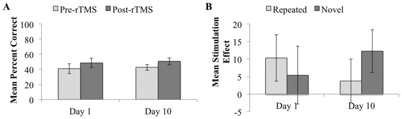
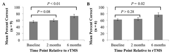
Methods: Nine patients received 10 days of 1-Hz rTMS (Monday through Friday for 2 weeks). We assessed naming performance before and immediately after stimulation on the first and last days of rTMS therapy, and then again at 2 and 6 months post-rTMS. A subset of six of these patients underwent functional magnetic resonance imaging pre-rTMS (baseline) and at 2 and 6 months post-rTMS.
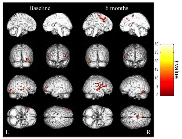
Results: Naming accuracy increased from pre- to post-rTMS on both the first and last days of treatment. We also found naming improvements long after rTMS, with the greatest improvements at 6 months post-rTMS. Long-lasting effects were associated with a posterior shift in the recruitment of the right inferior frontal gyrus: from the more anterior Brodmann area 45 to the more posterior Brodmann areas 6, 44, and 46. The number of left hemispheric regions recruited for naming also increased.
Conclusions: This study found that rTMS to the right hemisphere Broca area homologue confers long-lasting improvements in picture naming performance. The mechanism involves dynamic bilateral neural network changes in language processing, which take place within the right prefrontal cortex and the left hemisphere more generally.
Clinical trial registration: ClinicalTrials.gov (Identifier NCT00608582).
Conflict of interest statement
The authors declare no conflicts of interest.
Figures
References
-
- Barwood CH, Murdoch BE, Whelan BM, et al. The effects of low frequency repetitive transcranial magnetic stimulation (rTMS) and sham condition rTMS on behavioural language in chronic non-fluent aphasia: short term outcomes. NeuroRehabilitation. 2011;28:113–128. - PubMed
-
- Basso A, Gardelli M, Grassi MP, et al. The role of the right hemisphere in recovery from aphasia: two case studies. Cortex. 1989;25:555–566. - PubMed
-
- Berthier ML. Poststroke aphasia. Drugs Aging. 2005;22:163–182. - PubMed
-
- Chrysikou EG, Hamilton RH. Noninvasive brain stimulation in the treatment of aphasia: exploring interhemispheric relationships and their implications for neurorehabilitation. Restor Neurol Neurosci. 2011;29:375–394. - PubMed
MeSH terms
Associated data
Grants and funding
LinkOut - more resources
Full Text Sources
Other Literature Sources
Medical
