

Novel Image-Based Analysis for Reduction of Clinician-Dependent Variability in Measurement of the Corneal Ulcer Size
- PMID: 29256985
- PMCID: PMC5799030
- DOI: 10.1097/ICO.0000000000001488
Novel Image-Based Analysis for Reduction of Clinician-Dependent Variability in Measurement of the Corneal Ulcer Size
Abstract
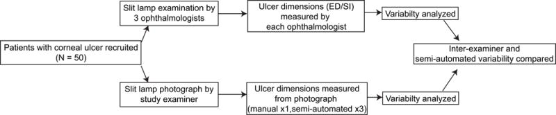
Purpose: To assess variability in corneal ulcer measurements between ophthalmologists and reduce clinician-dependent variability using semiautomated segmentation of the ulcer from photographs.
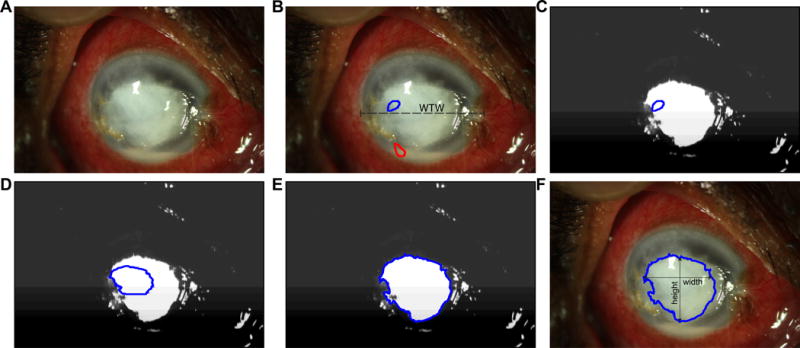
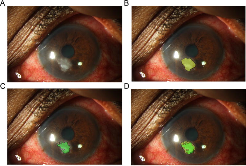
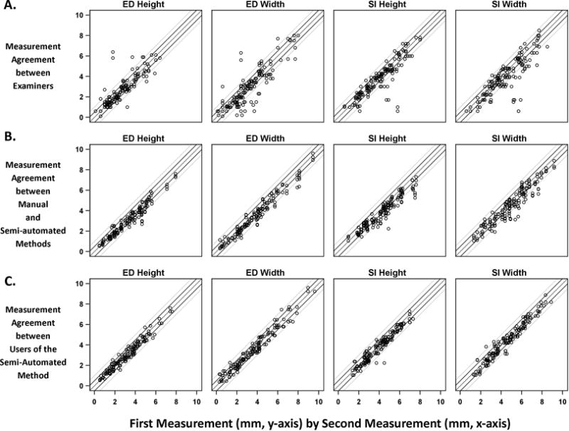
Methods: Three ophthalmologists measured 50 patients' eyes for epithelial defects (EDs) and the stromal infiltrate (SI) size using slit-lamp (SL) calipers. SL photographs were obtained. An algorithm was developed for semiautomatic segmenting of the ED and SI in the photographs. Semiautomatic segmentation was repeated 3 times by different users (2 ophthalmologists and 1 trainee). Clinically significant variability was assessed with intraclass correlation coefficients (ICCs) and the percentage of pairwise measurements differing by ≥0.5 mm. Semiautomatic segmentation measurements were compared with manual delineation of the image by a corneal specialist (gold standard) using Dice similarity coefficients.
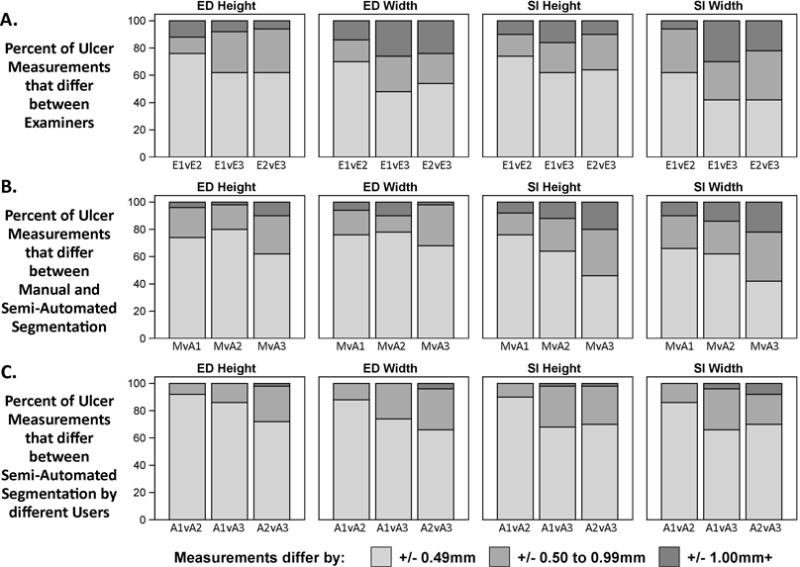
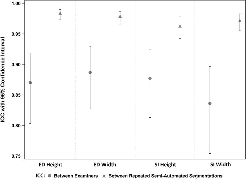
Results: Ophthalmologists' reliability in measurements by SL calipers had an ICC from 0.84 to 0.88 between examiners. Measurements by semiautomatic segmentation had an ICC from 0.96 to 0.98. SL measures of ulcers by clinical versus semiautomatic segmentation measures differed by ≥0.5 mm in 24% to 38% versus 8% to 28% (ED height); 30% to 52% versus 12% to 34% (ED width); 26% to 38% versus 10% to 32% (SI height); and 38% to 58% versus 14% to 34% (SI width), respectively. Average Dice similarity coefficients between manual and repeated semiautomatic segmentation ranged from 0.83 to 0.86 for the ED and 0.78 to 0.83 for the SI.
Conclusions: Variability exists when measuring corneal ulcers, even among ophthalmologists. Photography and computerized methods for quantifying the ulcer size could reduce variability while remaining accurate and impact quantitative measurement endpoints.
Conflict of interest statement
Figures
Similar articles
-
Clinical evaluation of corneal ulcer with a portable and smartphone-attachable slit lamp device: Smart Eye Camera.Sci Rep. 2025 Jan 24;15(1):3099. doi: 10.1038/s41598-025-87820-z. Sci Rep. 2025. PMID: 39856154 Free PMC article.
-
Evaluation of fungal keratitis using a newly developed computer program, Optscore, for grading digital corneal photographs.Ophthalmic Epidemiol. 2014 Feb;21(1):24-32. doi: 10.3109/09286586.2013.868003. Ophthalmic Epidemiol. 2014. PMID: 24467559 Free PMC article.
-
Semiautomatic quantification of carotid plaque volume with three-dimensional ultrasound imaging.J Vasc Surg. 2017 May;65(5):1407-1417. doi: 10.1016/j.jvs.2016.11.033. Epub 2017 Mar 6. J Vasc Surg. 2017. PMID: 28274755
-
Algorithm Variability in Quantification of Epithelial Defect Size in Microbial Keratitis Images.Cornea. 2020 May;39(5):628-633. doi: 10.1097/ICO.0000000000002258. Cornea. 2020. PMID: 31977729 Free PMC article.
-
Precision of Epithelial Defect Measurements.Cornea. 2017 Apr;36(4):419-424. doi: 10.1097/ICO.0000000000001148. Cornea. 2017. PMID: 28129296 Free PMC article.
Cited by
-
Artificial Intelligence in Cornea, Refractive Surgery, and Cataract: Basic Principles, Clinical Applications, and Future Directions.Asia Pac J Ophthalmol (Phila). 2021 Jul 1;10(3):268-281. doi: 10.1097/APO.0000000000000394. Asia Pac J Ophthalmol (Phila). 2021. PMID: 34224467 Free PMC article. Review.
-
Case report: Utilization of neutral density filters for densitometry analysis of dense corneal opacities.Am J Ophthalmol Case Rep. 2022 Jul 31;27:101672. doi: 10.1016/j.ajoc.2022.101672. eCollection 2022 Sep. Am J Ophthalmol Case Rep. 2022. PMID: 35966118 Free PMC article.
-
Open-Source Automatic Segmentation of Ocular Structures and Biomarkers of Microbial Keratitis on Slit-Lamp Photography Images Using Deep Learning.IEEE J Biomed Health Inform. 2021 Jan;25(1):88-99. doi: 10.1109/JBHI.2020.2983549. Epub 2021 Jan 5. IEEE J Biomed Health Inform. 2021. PMID: 32248131 Free PMC article.
-
Self-knowledge distillation-empowered directional connectivity transformer for microbial keratitis biomarkers segmentation on slit-lamp photography.Med Image Anal. 2025 May;102:103533. doi: 10.1016/j.media.2025.103533. Epub 2025 Mar 13. Med Image Anal. 2025. PMID: 40117989
-
Use of 'U-shaped tool for follow up of corneal ulcer cases in the COVID-19 pandemic.Indian J Ophthalmol. 2020 Oct;68(10):2199-2201. doi: 10.4103/ijo.IJO_1560_20. Indian J Ophthalmol. 2020. PMID: 32971640 Free PMC article.
References
-
- Bourne RR, Stevens GA, White RA, et al. Causes of vision loss worldwide, 1990–2010: a systematic analysis. Lancet Glob Health. 2013;1:e339–349. - PubMed
-
- Gonzales CA, Srinivasan M, Whitcher JP, et al. Incidence of corneal ulceration in Madurai district, South India. Ophthalmic Epidemiol. 1996;3:159–166. - PubMed
MeSH terms
Grants and funding
LinkOut - more resources
Full Text Sources
Other Literature Sources
Medical
