

THE INTEGRATIVE SURGICAL THEATER: Combining Intraoperative Optical Coherence Tomography and 3D Digital Visualization for Vitreoretinal Surgery in the DISCOVER Study
- PMID: 29256988
- PMCID: PMC6005714
- DOI: 10.1097/IAE.0000000000001999
THE INTEGRATIVE SURGICAL THEATER: Combining Intraoperative Optical Coherence Tomography and 3D Digital Visualization for Vitreoretinal Surgery in the DISCOVER Study
Abstract
Purpose: To evaluate the feasibility of integrating intraoperative optical coherence tomography (OCT) with a digital visualization platform for vitreoretinal surgery.
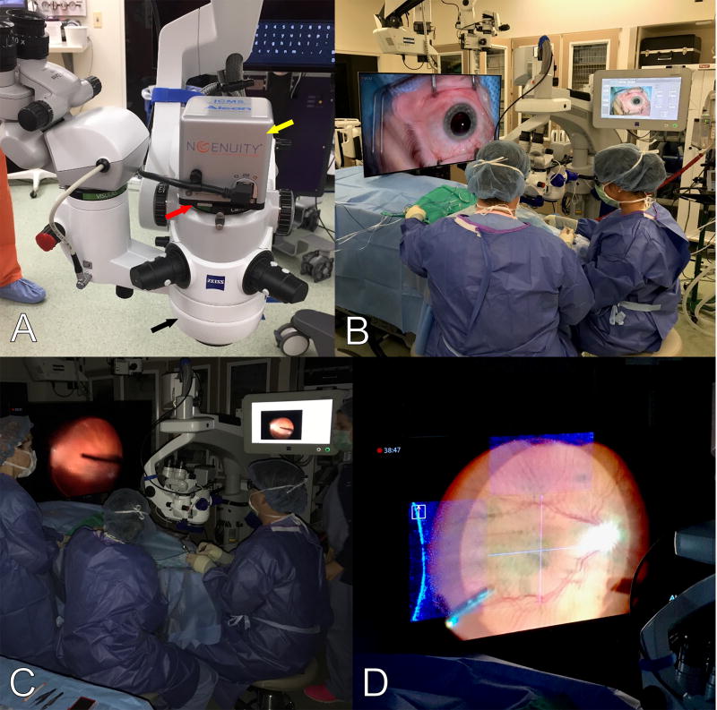
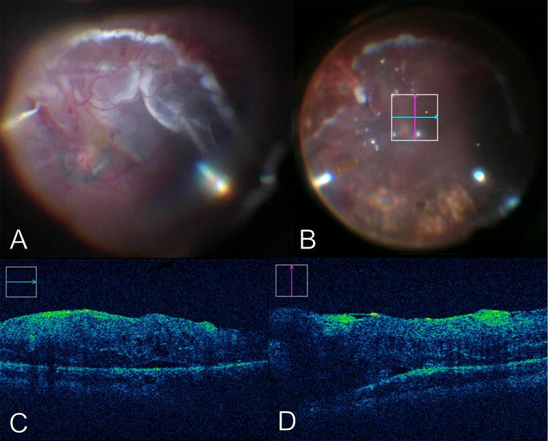
Methods: The DISCOVER study is a prospective study examining microscope-integrated intraoperative OCT across multiple prototypes and platforms. For this assessment, a microscope-integrated OCT platform was combined with a three-dimensional (3D) surgical visualization system to allow for digital display of the OCT data stream on the large immersive display. Intraoperative OCT scans were obtained at various surgical milestones that were directly overlaid to the surgical view in a 55-inch passive 3D 4K high-definition display. Surgeon feedback was obtained related to system performance and integration into the surgical procedures through a prespecified surgeon questionnaire.
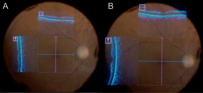
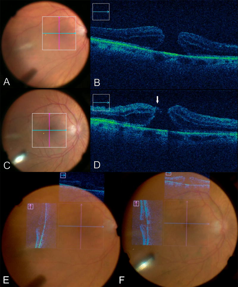
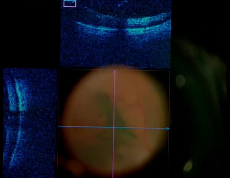
Results: Seven eyes of seven subjects were identified. Clinical diagnosis included epiretinal membrane (n = 3), macular hole (2), symptomatic vitreous opacity (1), and proliferative vitreoretinopathy (1). Optical coherence tomography images were successfully obtained and displayed on the 4K screen in all cases. Intraoperative OCT images facilitated identification of subtle retinal alterations. Surgeons reported that the 4K screen seemed to provide improved visualization of the OCT data stream compared with the semitransparent ocular view. Surgeons were able to examine the OCT data on the 4K screen without reverting to the external display system of the microscope. The system provided a uniform surgical visualization experience for both the surgeon and the assistant. In addition, the digital platform allowed all surgical personnel to simultaneously view both the OCT and the surgical field. All eyes underwent uneventful vitrectomy without reverting to the conventional microscope. No intraoperative adverse events occurred.
Conclusion: Integration of OCT into the digital visualization system may enable unique opportunities for surgeon feedback of intraoperative diagnostics. The overlay of the OCT data onto the 4K monitor seemed to provide excellent visualization of OCT details. Further research is needed to compare the conventional microscope-based approach to the digital 3D screen approach in regards to intraoperative OCT.
Figures
Similar articles
-
Pediatric Vitreoretinal Surgery and Integrated Intraoperative Optical Coherence Tomography.Dev Ophthalmol. 2021;61:15-25. doi: 10.1159/000511818. Epub 2021 Mar 1. Dev Ophthalmol. 2021. PMID: 33647898 Review.
-
Comparing Three-Dimensional Digitally Enabled Intraoperative OCT With Conventional Microscope-Integrated OCT in Vitreoretinal Surgery: A Post Hoc Analysis of the DISCOVER Study.Ophthalmic Surg Lasers Imaging Retina. 2024 May;55(5):270-277. doi: 10.3928/23258160-20240206-01. Epub 2024 Mar 1. Ophthalmic Surg Lasers Imaging Retina. 2024. PMID: 38648428
-
INTRAOPERATIVE OPTICAL COHERENCE TOMOGRAPHY DURING VITREORETINAL SURGERY FOR DENSE VITREOUS HEMORRHAGE IN THE PIONEER STUDY.Retina. 2015 Dec;35(12):2537-42. doi: 10.1097/IAE.0000000000000660. Retina. 2015. PMID: 26035403 Free PMC article.
-
An experimental and clinical study on the initial experiences of Brazilian vitreoretinal surgeons with heads-up surgery.Graefes Arch Clin Exp Ophthalmol. 2019 Mar;257(3):473-483. doi: 10.1007/s00417-019-04246-w. Epub 2019 Jan 15. Graefes Arch Clin Exp Ophthalmol. 2019. PMID: 30645695
-
Intraoperative Optical Coherence Tomography in Vitreoretinal Surgery.Semin Ophthalmol. 2019;34(4):312-316. doi: 10.1080/08820538.2019.1620811. Epub 2019 Jun 26. Semin Ophthalmol. 2019. PMID: 31240975 Review.
Cited by
-
Can the Three-Dimensional Heads-Up Display Improve Ergonomics, Surgical Performance, and Ophthalmology Training Compared to Conventional Microscopy?Clin Ophthalmol. 2021 Feb 18;15:679-686. doi: 10.2147/OPTH.S290396. eCollection 2021. Clin Ophthalmol. 2021. PMID: 33633441 Free PMC article.
-
Real-time visualization and interaction with static and live optical coherence tomography volumes in immersive virtual reality.Biomed Opt Express. 2018 May 30;9(6):2825-2843. doi: 10.1364/BOE.9.002825. eCollection 2018 Jun 1. Biomed Opt Express. 2018. PMID: 30258693 Free PMC article.
-
The DISCOVER Study 3-Year Results: Feasibility and Usefulness of Microscope-Integrated Intraoperative OCT during Ophthalmic Surgery.Ophthalmology. 2018 Jul;125(7):1014-1027. doi: 10.1016/j.ophtha.2017.12.037. Epub 2018 Mar 2. Ophthalmology. 2018. PMID: 29409662 Free PMC article.
-
Polarization-sensitive optical coherence tomography for renal tumor detection in ex vivo human kidneys.Opt Lasers Eng. 2024 Feb;173:107900. doi: 10.1016/j.optlaseng.2023.107900. Epub 2023 Oct 23. Opt Lasers Eng. 2024. PMID: 37982078 Free PMC article.
-
Preoperative imaging optimized for epiretinal membrane surgery.Int J Retina Vitreous. 2021 Apr 13;7(1):32. doi: 10.1186/s40942-021-00304-w. Int J Retina Vitreous. 2021. PMID: 33849642 Free PMC article.
References
-
- Ray R, et al. Intraoperative microscope-mounted spectral domain optical coherence tomography for evaluation of retinal anatomy during macular surgery. Ophthalmology. 2011;118:2212–2217. - PubMed
Publication types
MeSH terms
Grants and funding
LinkOut - more resources
Full Text Sources
Other Literature Sources
Medical
