. 2018 Jan;17(1):1651-1659.
doi: 10.3892/mmr.2017.8074. Epub 2017 Nov 15.

Protective effects of atorvastatin on cerebral vessel autoregulation in an experimental rabbit model of subarachnoid hemorrhage

Affiliations

Protective effects of atorvastatin on cerebral vessel autoregulation in an experimental rabbit model of subarachnoid hemorrhage

Jun-Hui Chen et al. Mol Med Rep. 2018 Jan.

Erratum in

Abstract

The aim of the present study was to assess the therapeutic effects of atorvastatin on cerebral vessel autoregulation and to explore the underlying mechanisms in a rabbit model of subarachnoid hemorrhage (SAH). A total of 48 healthy male New Zealand rabbits (weight, 2‑2.5 kg) were randomly allocated into SAH, Sham or SAH + atorvastatin groups (n=16/group). The Sham group received 20 mg/kg/d saline solution, whereas 20 mg/kg/d atorvastatin was administered to rabbits in the SAH + atorvastatin group following SAH induction. Changes in diameter, perimeter and basilar artery (BA) area were assessed and expression levels of the vasoactive molecules endothelin‑1 (ET‑1), von Willebrand factor (vWF) and thrombomodulin (TM) were measured. Neuronal apoptosis was analyzed 72 h following SAH by terminal deoxynucleotidyl-transferase‑mediated dUTP nick‑end labeling (TUNEL) staining. The mortality rate in the SAH group was 18.75, 25% in the SAH + atorvastatin treated group and 0% in the Sham group (n=16/group). The neurological score in the SAH + atorvastatin group was 1.75±0.68, which was significantly higher compared with the Sham group (0.38±0.49; P<0.05). The BA area in the SAH + atorvastatin group (89.6±9.11) was significantly lower compared with the SAH group (115.4±11.0; P<0.01). The present study demonstrated that SAH induction resulted in a significant increase in the diameter, perimeter and cross‑sectional area of the BA in the SAH + atorvastatin group. Administration of atorvastatin may significantly downregulate the expression levels of ET‑1, vWF and TM (all P<0.01) vs. sham and SAH groups. TUNEL staining demonstrated that neuronal apoptosis was remarkably reduced in the hippocampus of SAH rabbits following treatment with atorvastatin (P<0.05). Atorvastatin treatment may alleviate cerebral vasospasm and mediate structural and functional remodeling of vascular endothelial cells, in addition to promoting anti‑apoptotic signaling. These results provided supporting evidence for the use of atorvastatin as an effective and well‑tolerated treatment for SAH in various clinical settings and may protect the autoregulation of cerebral vessels.


Figures

Figure 1.
Figure 1.
Neurological scores in the experimental groups following SAH. The results indicated that the neurological score was significantly higher in SAH rats compared with rats in the Sham group at 72 h Atorvastatin reduced the neurological scores significantly than SAH group. *P<0.05 SAH + atorvastatin vs. SAH group. SAH, subarachnoid hemorrhage.
Figure 2.
Figure 2.
Effects of atorvastatin on the morphology of the BA (T score) in SAH rats. (A) Representative bright field images of the hematoxylin and eosin stained BA in the three experimental groups, procured with a light microscope; magnification, ×50. Diameter and cross-sectional area of the BA in the SAH group was smaller, whereas the BA wall became thicker compared with the Sham group. (B) Comparison of T values for each group. *P<0.01 vs. SAH. BA, basilar artery; SAH, subarachnoid hemorrhage.
Figure 3.
Figure 3.
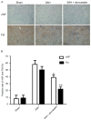
Immunohistochemical staining of brainstem for vWF and TM post-SAH. (A) Few vWF- and TM-positive regions were observed in the vessels in the Sham group, whereas positive staining increased in SAH-induced rats. The SAH-induced increases in expressions were notably reduced by atorvastatin treatment. (B) Quantitative analysis demonstrated a significant reduction in the percentage of positive-stained regions in the SAH + atorvastatin group compared with the SAH group. Magnification, ×50. Data are presented as the mean ± standard deviation; *P<0.01 and **P<0.01 vs. SAH group. SAH, subarachnoid hemorrhage; TM, thrombomodulin; vWF, von Willebrand factor.
Figure 4.
Figure 4.
TUNEL staining of hippocampus following SAH induction. (A) Few apoptotic cells were observed in Sham group rabbits. TUNEL-positive staining was markedly increased in SAH rabbits, which was notably reduced by atorvastatin treatment. (B) Quantitative analysis demonstrated a significant increase in the number of apoptotic cells (number/mm2) by SAH induction compared with the number of apoptotic cells in the Sham group; *P<0.05. A significant reduction in number of apoptotic-positive cells was observed in the SAH + atorvastatin group compared with the SAH group; #P<0.05. SAH, subarachnoid hemorrhage; TUNEL, terminal deoxynucleotidyl-transferase-mediated dUTP nick-end labeling.
Figure 5.
Figure 5.
ELISA-based examination of the effects of atorvastatin treatment on the concentration of ET-1 in the plasma of the rabbits at 72 h following SAH. Graph demonstrating the protein concentration of ET-1 in the three groups (n=8/group). Data are presented as the mean ± standard deviation. ET-1 was significantly increased in SAH group compared with Sham group at 72 h; *P<0.01. Atorvastatin treatment significantly decreased the SAH-induced ET-1 expression compared with the SAH group; #P<0.01. ET-1, endothelin 1; SAH, subarachnoid hemorrhage.
Figure 6.
Figure 6.
mRNA expression levels of vWF and TM in brain tissues were detected by reverse transcription-quantitative polymerase chain reaction. Data are presented as the mean ± standard deviation; n=8/group. The mRNA expression levels of vWF and TM were significantly increased in SAH group compared with the Sham group at 72 h in the plasma; *P<0.01; Atorvastatin treatment significantly decreased the mRNA expression levels of vWF and TM compared with the SAH group; #P<0.01. SAH, subarachnoid hemorrhage; TM, thrombomodulin; vWF, von Willebrand factor.
Figure 7.
Figure 7.
Caspase-3 protein expression in the hippocampus. (A) Representative western blot of Caspase-3 protein expression. (B) Densitometric analysis of Caspase-3 protein expression. Data are presented as the mean ± standard deviation. Caspase-3 protein expression in the Sham group was significantly lower than SAH group and SAH + atorvastatin group (n=8/group, #P<0.01). Caspase-3 expression levels were significantly decreased in the SAH + atorvastatin group vs. the SAH group; *P<0.05. Equal protein loading was confirmed by intracellular GAPDH protein expression. SAH, subarachnoid hemorrhage.

References

    1. Komotar RJ, Schmidt JM, Starke RM, Claassen J, Wartenberg KE, Lee K, Badjatia N, Connolly ES, Jr, Mayer SA. Resuscitation and critical care of poor-grade subarachnoid hemorrhage. Neurosurgery. 2009;64:397–411. doi: 10.1227/01.NEU.0000338946.42939.C7. - DOI - PubMed
    1. Rosengart AJ, Schultheiss KE, Tolentino J, Macdonald RL. Prognostic factors for outcome in patients with aneurysmal subarachnoid hemorrhage. Stroke. 2007;38:2315–2321. doi: 10.1161/STROKEAHA.107.484360. - DOI - PubMed
    1. Steiner T, Juvela S, Unterberg A, Jung C, Forsting M, Rinkel G. European Stroke Organization: European Stroke Organization guidelines for the management of intracranial aneurysms and subarachnoid haemorrhage. Cerebrovasc Dis. 2013;35:93–112. doi: 10.1159/000346087. - DOI - PubMed
    1. Budohoski KP, Czosnyka M, Kirkpatrick PJ, Smirlewski P, Steiner LA, Pickard JD. Clinical relevance of cerebral autoregulation following subarachnoid haemorrhage. Nat Rev Neurol. 2013;9:152–163. doi: 10.1038/nrneurol.2013.11. - DOI - PubMed
    1. Laskowitz DT, Kolls BJ. Neuroprotection in subarachnoid hemorrhage. Stroke. 2010;41(10 Suppl):S79–S84. doi: 10.1161/STROKEAHA.110.595090. - DOI - PMC - PubMed

MeSH terms