Comparative Study
. 2018 Feb;17(2):2879-2884.
doi: 10.3892/mmr.2017.8239. Epub 2017 Dec 11.

N‑cadherin attenuates nucleus pulposus cell senescence under high‑magnitude compression

Affiliations
Comparative Study

N‑cadherin attenuates nucleus pulposus cell senescence under high‑magnitude compression

Ming Niu et al. Mol Med Rep. 2018 Feb.

Abstract

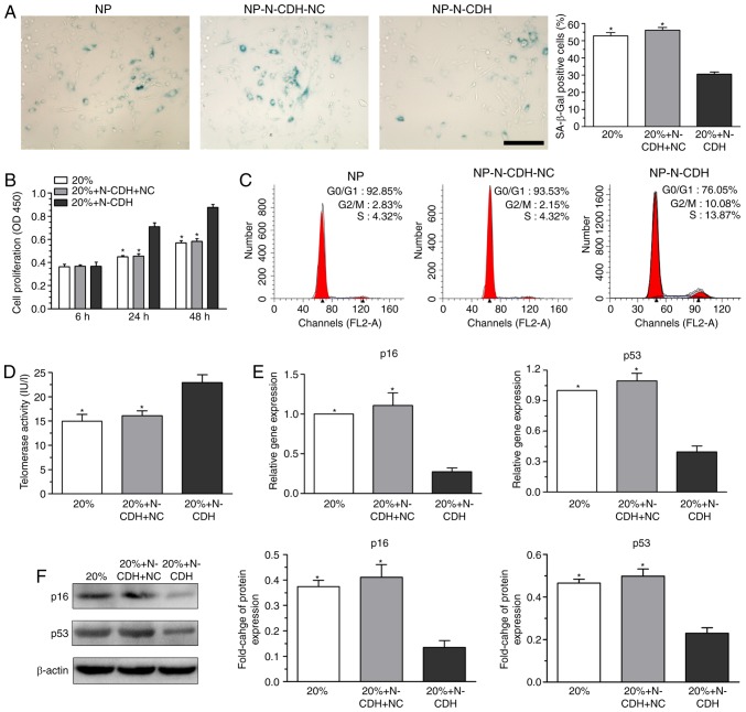
Mechanical compression is important in disc degeneration. N-cadherin (N-CDH)-mediated signaling contributes to the maintenance of the normal nucleus pulposus (NP) cell phenotype and NP matrix biosynthesis. Our preliminary study demonstrated that a high‑magnitude compression (20% deformation) promotes NP cell senescence in a three‑dimensional scaffold culture system. The aim of the present study was to investigate whether N‑CDH‑mediated signaling alleviates NP cell senescence under the above‑mentioned high‑magnitude compression. NP cells were transfected with recombinant lentiviral vectors to enhance N‑CDH expression. All the transfected or un‑transfected NP cells were seeded into the scaffolds and subjected to 20% deformation at a frequency of 1.0 Hz for 4 h once per day for 5 days. Results indicated that N‑CDH overexpressed NP cells exhibited decreased senescence‑associated β‑galactosidase activity and downregulated expression levels of senescence‑associated markers (p16 and p53). Furthermore, the N‑CDH overexpressed NP cells exhibited increased cell proliferation potency, telomerase activity and matrix biosynthesis compared with NP cells without N‑CDH overexpression under high‑magnitude compression. Thus, N‑CDH‑mediated signaling contributes to the attenuation of NP cell senescence under high‑magnitude compression.


Figures

Figure 1.
Figure 1.
Image of the substance exchanger-based perfusion bioreactor. 1, medium reservoir; 2, substance exchanger; 3, peristaltic pump; 4, tissue culture chamber; 5, pH, PO2 and PCO2 sensor.
Figure 2.
Figure 2.
Verification of N-CDH overexpression in NP cells. (A) Successfully transfected NP cells were tagged with green fluorescent protein. Magnification, ×200. (B) Quantitative polymerase chain reaction and western blotting assays indicated that N-CDH expression levels were upregulated in NP cells following incubation with recombinant lentiviral vectors. (C) N-CDH expression was increased in NP cells under the high-magnitude compression (20% compressive deformation) following N-CDH overexpression. Data are expressed as the mean ± standard deviation (n=3). *P<0.05, indicates a significant difference between two groups. N-CDH, N-cadherin; NP, nucleus pulposus; NC, negative control.
Figure 3.
Figure 3.
N-CDH overexpression attenuated NP cell senescence under the high-magnitude compression (20% compressive deformation). (A) SA-β-Gal activity analysis (scale bar, 100 µm). (B) Cell proliferation analysis. (C) Cell cycle analysis. (D) Telomerase activity measurement. (E) Quantitative polymerase chain reaction analysis of gene expression of senescence markers (p16 and p53). (F) Western blot analysis of protein expression levels of senescence markers (p16 and p53). Data are expressed as the mean ± standard deviation (n=3). *P<0.05, indicates a significant difference between two groups. N-CDH, N-cadherin; NP, nucleus pulposus; NC, negative control.
Figure 4.
Figure 4.
N-CDH overexpression upregulated the expression levels of NP matrix macromolecules (aggrecan and collagen II) under the high-magnitude compression (20% compressive deformation). (A and B) Quantitative polymerase chain reaction and western blot analysis of the expression levels of aggrecan and collagen II, respectively. Data are expressed as the mean ± standard deviation (n=3). *P<0.05, indicates a significant difference between two groups. N-CDH, N-cadherin; NP, nucleus pulposus; NC, negative control.

References

    1. Chen JW, Ni BB, Li B, Yang YH, Jiang SD, Jiang LS. The responses of autophagy and apoptosis to oxidative stress in nucleus pulposus cells: Implications for disc degeneration. Cell Physiol Biochem. 2014;34:1175–1189. doi: 10.1159/000366330. - DOI - PubMed
    1. Yang SD, Ma L, Gu TX, Ding WY, Zhang F, Shen Y, Zhang YZ, Yang DL, Zhang D, Sun YP, Song YL. 17β-Estradiol protects against apoptosis induced by levofloxacin in rat nucleus pulposus cells by upregulating integrin α2β1. Apoptosis. 2014;19:789–800. doi: 10.1007/s10495-014-0965-4. - DOI - PubMed
    1. Yang SD, Ma L, Yang DL, Ding WY. Combined effect of 17β-estradiol and resveratrol against apoptosis induced by interleukin-1β in rat nucleus pulposus cells via PI3K/Akt/caspase-3 pathway. PeerJ. 2016;4:e1640. doi: 10.7717/peerj.1640. - DOI - PMC - PubMed
    1. Yang SD, Yang DL, Sun YP, Wang BL, Ma L, Feng SQ, Ding WY. 17β-estradiol protects against apoptosis induced by interleukin-1β in rat nucleus pulposus cells by down-regulating MMP-3 and MMP-13. Apoptosis. 2015;20:348–357. doi: 10.1007/s10495-015-1086-4. - DOI - PubMed
    1. Lee CR, Iatridis JC, Poveda L, Alini M. In vitro organ culture of the bovine intervertebral disc: Effects of vertebral endplate and potential for mechanobiology studies. Spine (Phila Pa 1976) 2006;31:515–522. doi: 10.1097/01.brs.0000201302.59050.72. - DOI - PMC - PubMed

Publication types