Review
. 2017 Dec 16;18(12):2733.
doi: 10.3390/ijms18122733.

Molecular Biology of Prune Dwarf Virus-A Lesser Known Member of the Bromoviridae but a Vital Component in the Dynamic Virus-Host Cell Interaction Network

Affiliations
Review

Molecular Biology of Prune Dwarf Virus-A Lesser Known Member of the Bromoviridae but a Vital Component in the Dynamic Virus-Host Cell Interaction Network

Edmund Kozieł et al. Int J Mol Sci. .

Abstract

Prune dwarf virus (PDV) is one of the members of Bromoviridae family, genus Ilarvirus. Host components that participate in the regulation of viral replication or cell-to-cell movement via plasmodesmata are still unknown. In contrast, viral infections caused by some other Bromoviridae members are well characterized. Bromoviridae can be distinguished based on localization of their replication process in infected cells, cell-to-cell movement mechanisms, and plant-specific response reactions. Depending upon the genus, "genome activation" and viral replication are linked to various membranous structures ranging from endoplasmic reticulum, to tonoplast. In the case of PDV, there is still no evidence of natural resistance sources in the host plants susceptible to virus infection. Apparently, PDV has a great ability to overcome the natural defense responses in a wide spectrum of plant hosts. The first manifestations of PDV infection are specific cell membrane alterations, and the formation of replicase complexes that support PDV RNA replication inside the spherules. During each stage of its life cycle, the virus uses cell components to replicate and to spread in whole plants, within the largely suppressed cellular immunity environment. This work presents the above stages of the PDV life cycle in the context of current knowledge about other Bromoviridae members.

Keywords: Bromoviridae; Prune dwarf virus; plant defense response; plant–virus interactions; replication process; systemic and local movement.


Conflict of interest statement

The authors declare no conflict of interest.

Figures

Figure 1
Figure 1
Genome structure of Prune dwarf virus (PDV). Scheme presenting the individual open reading frames (dark blue) and proteins (different colors) encoded by PDV RNAs. Encoded proteins: P1 with methyltransferase and helicase domains (yellow), P2—polymerase (red), MP—movement protein (grey), CP—coat protein (green). ORF—open reading frame, RBD—RNA binding domain, HR—hydrophobic region. Scheme of genome prepared on the basis of the data in Bujarski et al. [1].
Figure 2
Figure 2
Immunofluorescent localization of P1 protein (replicase) in the tobacco leaf tissue of Samsun variety. (A) Immunofluorescent visualization of the epitopes of P1 protein (green, marked with *) in palisade and spongy mesophyll cells (cross-section of tobacco leaf blade); (B) Epitopes of P1 protein (*) visible in parenchyma and necrotic altered phloem; (C) Cross section of tobacco leaf 15 days after inoculation with buffer. No locations of P1 protein epitopes. Abbreviations: Ep—epidermis, PMe—palisade mesophyll, SMe—spongy mesophyll, Pa—parenchyma, X—tracheal element, Ph—phloem, Ne—necrosis. Kozieł et al. [32] modified.
Figure 3
Figure 3
Immunogold localization of P1 protein (replicase) in mesophyll and phloem cells of tobacco leaf of Samsun variety 15 days after inoculation with PDV. (A) Colloidal gold particles associated with P1 epitope (*) in vacuoles, vesicles and chloroplasts of palisade parenchyma cell; Bar 1µm (B) Gold particles in palisade parenchyma cell (*) in chloroplast, vacuole, and in vicinity of mitochondria with electron-translucent area; Bar 1µm (C) Gold particles (*) in parenchyma cell tonoplast and in membranes of spherules. The white framed area is enlarged in (D); Bar 1µm (D) Enlarged fragment with spherules in the white frame from (C); Bar 0,5 µm (E) Epitopes of P1 protein (*) in vacuoles of phloem parenchyma and companion cells, and inside sieve tubes. Viral particles in companion cell; Bar 2µm (F) Colloidal gold particles (*) in companion cell vacuoles; Bar 2 µm (G) Control tobacco plant (mock-inoculated) phloem without of P1 localization Bar 1µm. Abbreviations: CW—cell wall, Ch—chloroplast, ER—endoplasmic reticulum, V—vacuole, vs—vesicle, M—mitochondrion, Sp—spherule, PD—plasmodesmata, SE—sieve tube, CC—companion cell, PP—phloem parenchyma, VP—viral particles, N—nucleus. Kozieł et al. [32] modified.
Figure 4
Figure 4
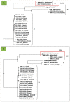
Phylogenetic comparison of amino acid sequences of the MP and RNA binding domain of PDV with several members of Bromoviridae family: (A) MP sequences for the studied groups of virus isolates. The highest similarity (maximum likelihood) between PDV and AMV marked in red frame (34%) (modified [36]); (B) RNA binding domain of the MPs. The highest similarity (maximum likelihood) between PDV and AMV marked within the red frame (about level 40%), Kozieł et al. [36] modified. On (A)and (B): I- group of analyzed PDV isolates sequences of movement protein, II- group of analyzed Brome mosaic virus (BMV) and Cucumber mosaic virus (CMV) isolates sequences of movement protein, III- group of analyzed AMV and Cowpea chlorotic mottle virus (CCMV) isolates sequences of movement protein.
Figure 5
Figure 5
Immunofluorescent localization of coat protein in tobacco leaf of Samsun variety 15 days after inoculation with PDV. (A) The epitopes of coat protein (*) in phloem cells, palisade, and spongy mesophyll. Disintegration of spongy mesophyll cells observed (arrow); (B) The epitopes of coat protein (*) in necrotic phloem cells. Visible cell wall invagination of parenchyma cells (arrow); (C) The epitopes of coat protein (*) in the spongy mesophyll cell and tracheal element; (D) Fragments of leaf blades inoculated only with buffer after 15 days. No CP epitopes observed. Abbreviations: Ep—epidermis, PMe—palisade mesophyll, SMe—spongy mesophyll, X—tracheal element of xylem, Ph—phloem, Ne—necrosis. Kozieł et al. [32] modified.
Figure 6
Figure 6
Immunogold localization of PDV coat protein (CP) in the tissues of the tobacco leaf blade, Samsun variety, 15 days post inoculation with PDV. (A) Labeling is observed in nucleus (N) and on the endoplasmic reticulum (ER) surface (*); (B) Colloidal gold particles (*) in vacuole of mesophyll cell. Labeling observed in cytoplasm (arrow) and by plasmodesmata; (C) Colloidal gold particles (*) in the companion cell protoplast. Labeling is observed both in vesicles and in the vacuole; (D) Colloidal gold particles (*) in the spherules’ membranes in a parenchyma cell; (E) Control tobacco plant phloem without of CP localization. Abbreviations: CW—cell wall, Ch—chloroplast, M—mitochondrion, ER—endoplasmic reticulum, VP—viral particles, vs—vesicle, SE—sieve tube, Sp—spherule, PD−plasmodesmata; St—starch−, Bars 1 µm.Kozieł et al. [32] modified.
Figure 7
Figure 7
Three-dimensional (3D) model structure of the PDV replicase (P1) protein, considering the surface of both N- and C-terminal domain, and the transmembrane domain (modified Kozieł et al., 2017). (A) The colors show the particular elements of the secondary structure, as follows. Green indicates the fragments of straight polypeptide chain, blue—α-helical fragments, yellow—β-card fragments; orange—the frame of methyltransferase domain, white—the frame of helicase domain. The central buckle depicts the helical region between both domains. The protein framework of the 3D structure is marked in gray; (B) Magnification of the methyltransferase domain from Figure 6a. Gray indicates protein surface; (C) Magnification of the helicase domain (from Figure 6A) rotated 90 degrees vis-à-vis the vertical axis, with the region used for immunolocalization of P1 marked in red. Purple indicates the predicted transmembrane domain. The area of the general framework of the helicase C-terminal domain is represented by the gray color. Kozieł et al. [32], modified.
Figure 8
Figure 8
(A) 3D visualization of a model of the PDV replication complex that assembles during the first steps of viral RNA replication; (A) P1 protein (of the replicase complex), marked by blue color, as it anchors to the tonoplast membrane; (B) P2 protein (RNA dependent RNA polymerase) 3D surface structure, as marked by green color. (C) Fully assembled replication complex, consists of P1 protein (in blue) attached to tonoplast membrane and P2 protein (in green), that is attached to P1 between its both domains. This computer-generated model was generated by using THMM program, in ΔG Prediction Server 1.0, and in AIDA server. To display the results of these calculations, the CELLmicrocosmos Membrane Editor was utilized.
Figure 9
Figure 9
Model of the pathogen life cycle, from replication to cell-to-cell transport, by compiling both TMV and CCMV as the examples of viral RNA (vRNA) replicated at the endoplasmic reticulum (ER), and then transported as a complex with movement protein (MP) along actin microfilaments (MF) to the plasmodesmata (PD) region. Simultaneously, MP is also transported alongside microtubules to PD. MP modifies the size exclusion limit (SEL) of PD. Frame with *TMV, CCMV replication sites of vRNA. Frame with ***TMV, CCMV complexes of vRNA and MP, transported with help of MF.
Figure 10
Figure 10
Model of a viral pathogen life cycle, from replication to cell-to-cell transport, based upon CMV characteristics. Viral RNA (vRNA) replication occurs at the endoplasmatic reticulum (ER) and the RNA is transported along actin microfilaments (MF) to plasmodesmata as a complex consisting of coat protein (CP) and movement protein (MP). MP modifies the size exclusion limit of plasmodesmata. Frame with *CMV-replication sites of vRNA. Frame with ***CMV vRNA, MP and CP complex transported with help of MF-route (mechanism) of cell-to-cell transport.
Figure 11
Figure 11
A model of PDV life cycle including replication and cell-to-cell transport, based upon research data for BMV and AMV. The BMV replication complex (P1 and P2) is assembled inside the spherules of the endoplasmic reticulum (ER) membranes. Viral RNA of BMV is encapsidated inside virion shells composed of CP molecules. In contrast, in the case of AMV and likely of PDV, the assembly of the replication complex and RNA replication are connected with the tonoplast membrane. As for BMV, the AMV and PDV RNAs are encapsidated with CP molecules. Thereafter, the assembled BMV or AMV (and likely PDV) virions are transported inside the microtubules (MT) that were modified with MP molecules. MP both change the PD size exclusion limit (SEL), but also destroy desmotubule structures inside PD. Frame with *BMV—replication sites of vRNA. Frame with **AMV, PDV replication sites of vRNA. Frame with ***BMV, AMV, PDV viral particles transported with help of MP an MT—in the case of PDV, probably route (mechanism) of cell-to-cell transport.

Similar articles

Cited by

References

    1. Bujarski J.J., Figlerowicz M., Gallitelli D., Roossinck M.J., Scott S.W. Family Bromoviridae. In: King A.M.Q., Adams M.J., Carstens E.B., Lefkowitz E.J., editors. Virus Taxonomy: Classification and Nomenclature of Viruses-Ninth Report of the International Committee on Taxonomy of Viruses. 1st ed. Elsevier Academic Press; San Diego, CA, USA: 2012. pp. 972–976.
    1. Brunt H.A., Crabtree K., Dallawitz M.J., Gibs A.J., Watson L. Viruses of Plants. 1st ed. CAB International UK; Wallingford, UK: 1996.
    1. Codoñer F.M., Cuevas J.M., Sanchez-Navarro J.A., Pallas V., Elena S.F. Molecular evolution of the plant virus family Bromoviridae based on RNA3-encoded proteins. J. Mol. Evol. 2005;61:697–705. doi: 10.1007/s00239-005-0021-7. - DOI - PubMed
    1. Codoñer F.M., Fares M.A., Elena S.F. Adaptive covariation between the coat and movement proteins of Prunus necrotic ringspot virus. J. Virol. 2006;80:5833–5840. doi: 10.1128/JVI.00122-06. - DOI - PMC - PubMed
    1. International Committee on Taxonomy of Viruses (ICTV) Official Website. [(accessed on 16 November 2017)]; Available online: https://talk.ictvonline.org/ictv-reports/ictv_9th_report/positive-sense-....

MeSH terms

Supplementary concepts

LinkOut - more resources