. 2017 Dec 18;22(12):2251.
doi: 10.3390/molecules22122251.

The Benefits of the Citrus Flavonoid Diosmin on Human Retinal Pigment Epithelial Cells under High-Glucose Conditions

Affiliations

The Benefits of the Citrus Flavonoid Diosmin on Human Retinal Pigment Epithelial Cells under High-Glucose Conditions

Wayne Young Liu et al. Molecules. .

Abstract

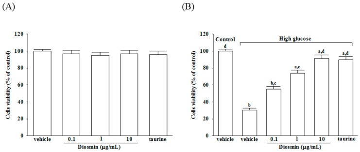
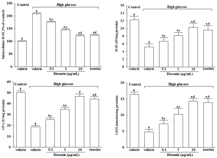
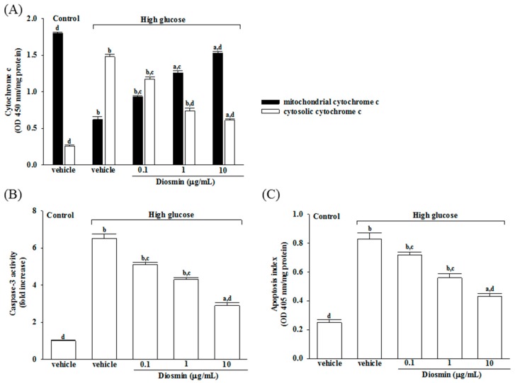
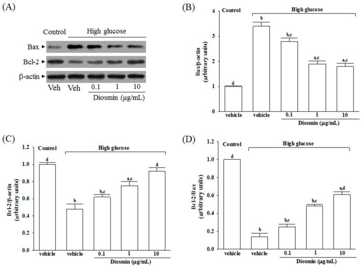
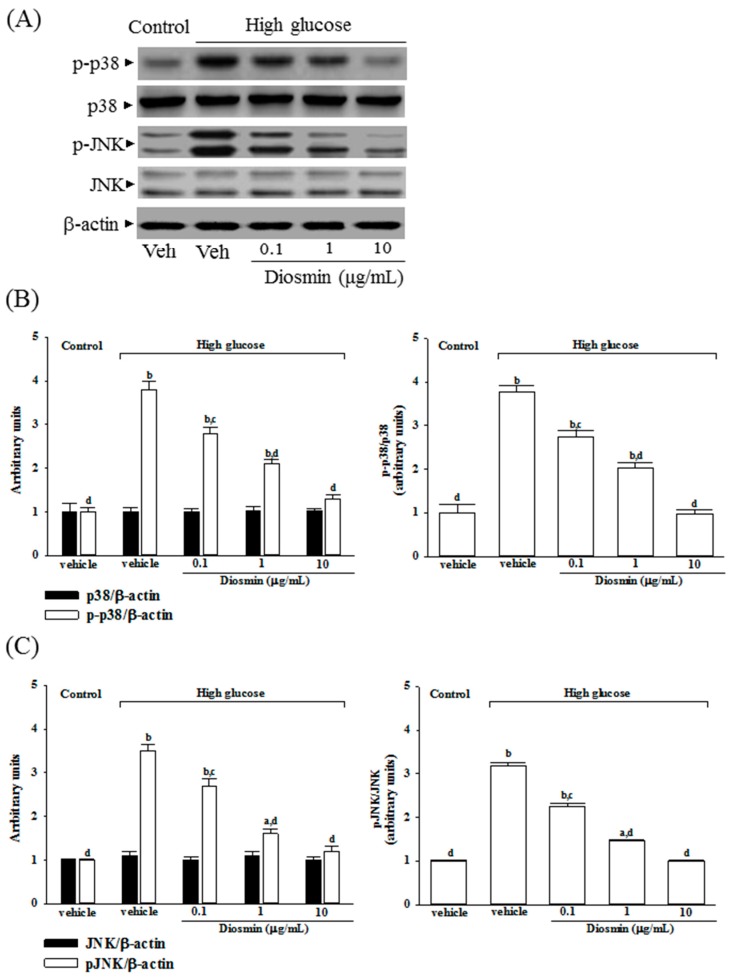
We investigate diosmin for its effect on the ARPE-19 human retinal pigment epithelial cells exposed to high glucose, a model of diabetic retinopathy (DR). After incubation for 4 days with a normal (5 mmol/L) concentration of D-glucose, ARPE-19 cells were exposed separately to normal or high concentrations of D-glucose (30 mmol/L) with or without diosmin at different concentrations (0.1, 1, 10 μg/mL) for another 48 h. Next, we assessed cell viability, reactive oxygen species (ROS) generation and antioxidant enzyme activities. In order to examine the underlying molecular mechanisms, we meanwhile analyzed the expressions of Bax, Bcl-2, total and phosphorylated JNK and p38 mitogen-activated protein kinase (MAPK). Diosmin dose dependently enhanced cell viability following high glucose treatment in ARPE-19 cells. The activities of superoxide dismutase and glutathione peroxidase, as well as the levels of reduced glutathione were decreased, while it was observed that levels of ROS in high glucose cultured ARPE-19 cells increased. High glucose also disturbed Bax and Bcl-2 expression, interrupted Bcl-2/Bax balance, and triggered subsequent cytochrome c release into the cytosol and activation of caspase-3. These detrimental effects were ameliorated dose dependently by diosmin. Furthermore, diosmin could abrogate high glucose-induced apoptosis as well as JNK and P38 MAPK phosphorylation in ARPE-19 cells. Our results suggest that treatment ARPE-19 cells with diosmin halts hyperglycemia-mediated oxidative damage and thus this compound may be a candidate for preventing the visual impairment caused by DR.

Keywords: MAPK; ROS; diabetic retinopathy; diosmin; high glucose; human retinal pigment epithelial cells.


Conflict of interest statement

The authors declare no conflicts of interest in relation to this work.

Figures

Figure 1
Figure 1
Cytotoxicity of diosmin to ARPE-19 cells. Normal glucose (A) or high glucose (B) cultured ARPE-19 cells were treated with or without different concentrations of diosmin (0.1, 1, 10 μg/mL) or taurine (1 mmol/L) for 48 h. Cell viability was conducted by MTT assay. The results are presented as the mean ± SD of five independent experiments. a p < 0.05 and b p < 0.01 compared with normal-glucose vehicle-treated group (control), respectively. c p < 0.05 and d p < 0.01 compared with high-glucose vehicle-treated group, respectively.
Figure 2
Figure 2
Effect of diosmin on the ROS generation and the antioxidant biomolecules in high glucose cultured ARPE-19 cells. The results are presented as the mean ± SD of five independent experiments in each column. a p < 0.05 and b p < 0.01 compared with normal-glucose vehicle-treated group (control), respectively. c p < 0.05 and d p < 0.01 compared with high-glucose vehicle-treated group, respectively.
Figure 3
Figure 3
Effects of diosmin on high glucose-induced mitochondrial dysfunction in ARPE-19 cells. (A) Levels of cytochrome c in the cytosolic fraction or mitochondrial fractions were determined by ELISA kit; (B) The activity of caspase-3 in the cell lysate was measured using a colorimetric caspase-3 assay kit; (C) Apoptosis index was measured by detection of DNA fragmentation with the Cell death detection ELISA kit. Each column represents means ± SD (n = 5 per group). a p < 0.05 and b p < 0.01 compared with normal-glucose vehicle-treated group (control), respectively. c p < 0.05 and d p < 0.01 compared with high-glucose vehicle-treated group, respectively.
Figure 4
Figure 4
Effect of diosmin on Bax and Bcl-2 expression in high glucose cultured ARPE-19 cells. (A) The photographs were representatives the Western blots for Bax and Bcl-2 protein. Five western blots per group were experimented; (B) Relative intensities of Bax protein (fold to β-actin); (C) Relative intensities of Bcl-2 protein (fold to β-actin); (D) Ratio of the relative intensities of Bcl-2 to Bax (Bcl-2/Bax). Each column represents means ± SD (n = 5 per group). a p < 0.05 and b p < 0.01 compared with normal-glucose vehicle-treated group (control), respectively. c p < 0.05 and d p < 0.01 compared with high-glucose vehicle-treated group, respectively.
Figure 5
Figure 5
Effect of diosmin on p38 and JNK activation in high glucose cultured ARPE-19 cells. (A) The photographs were representatives the western blots for p-p38, p38, p-JNK, JNK and β-actin. Five western blots per group were experimented; (B) The relative intensities of phosphorylated and total p38 protein (fold to β-actin) were determined and the p-p38/p38 ratio was calculated; (C) The relative intensities of phosphorylated and total JNK protein (fold to β-actin) was determined and the p-JNK/JNK ratio was calculated. Each column represents means ± SD (n = 5 per group). a p < 0.05 and b p < 0.01 compared with normal-glucose vehicle-treated group (control), respectively. c p < 0.05 and d p < 0.01 compared with high-glucose vehicle-treated group, respectively.

References

    1. Chen Y.H., Chou H.C., Lin S.T., Chen Y.W., Lo Y.W., Chan H.L. Effect of high glucose on secreted proteome in cultured retinal pigmented epithelium cells: Its possible relevance to clinical diabetic retinopathy. J. Proteom. 2012;77:111–128. doi: 10.1016/j.jprot.2012.07.014. - DOI - PubMed
    1. Strauss O. The retinal pigment epithelium in visual function. Physiol. Rev. 2005;85:845–881. doi: 10.1152/physrev.00021.2004. - DOI - PubMed
    1. Desjardins D.M., Yates P.W., Dahrouj M., Liu Y., Crosson C.E., Ablonczy Z. Progressive early breakdown of retinal pigment epithelium function in hyperglycemic rats. Investig. Ophthalmol. Vis. Sci. 2016;57:2706–2713. doi: 10.1167/iovs.15-18397. - DOI - PMC - PubMed
    1. Koopman W.J., Nijtmans L.G., Dieteren C.E., Roestenberg P., Valsecchi F., Smeitink J.A., Willems P.H. Mammalian mitochondrial complex I: Biogenesis, regulation, and reactive oxygen species generation. Antioxid. Redox Signal. 2010;12:1431–1470. doi: 10.1089/ars.2009.2743. - DOI - PubMed
    1. Jin G.F., Hurst J.S., Godley B.F. Rod outer segments mediate mitochondrial DNA damage and apoptosis in human retinal pigment epithelium. Curr. Eye Res. 2001;23:11–19. doi: 10.1076/ceyr.23.1.11.5423. - DOI - PubMed

MeSH terms