. 2017 Dec 18;9(12):388.
doi: 10.3390/v9120388.

Stress Granule-Inducing Eukaryotic Translation Initiation Factor 4A Inhibitors Block Influenza A Virus Replication

Affiliations

Stress Granule-Inducing Eukaryotic Translation Initiation Factor 4A Inhibitors Block Influenza A Virus Replication

Patrick D Slaine et al. Viruses. .

Abstract

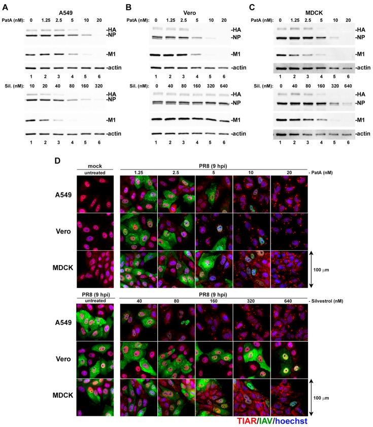
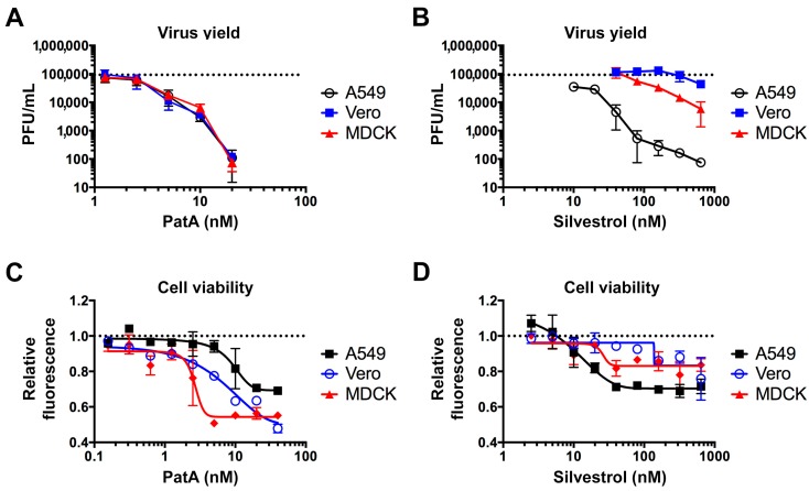
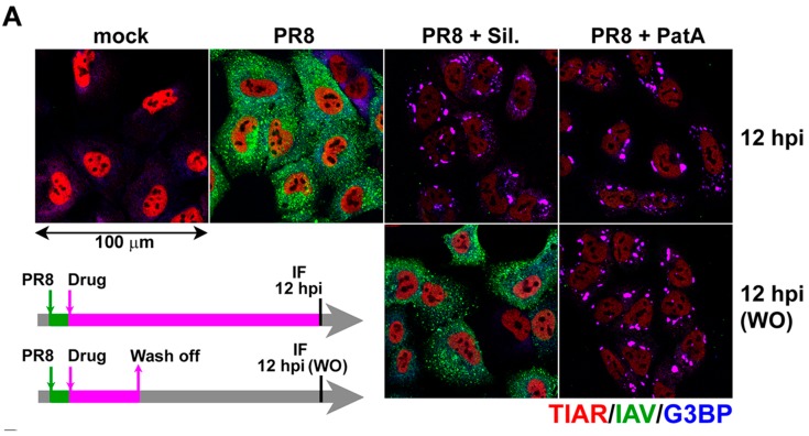
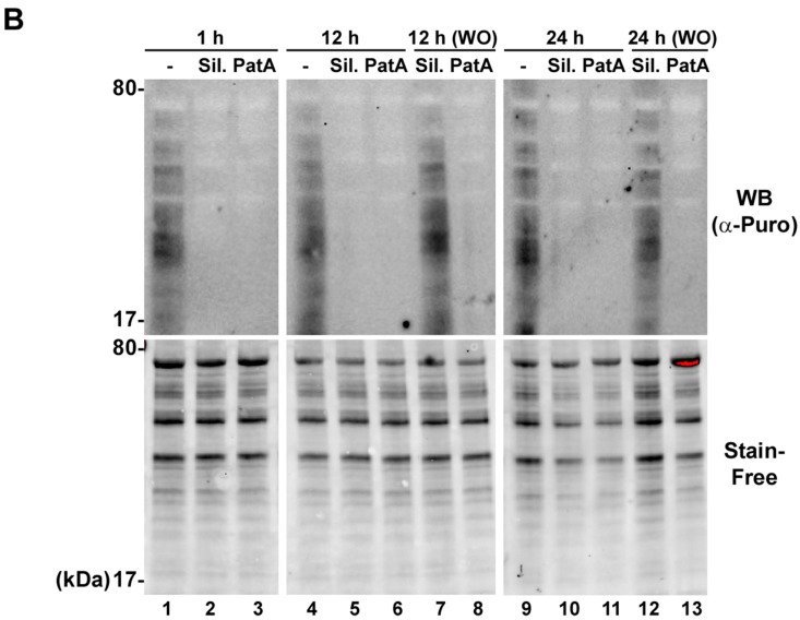
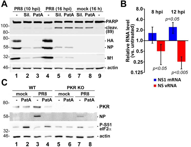
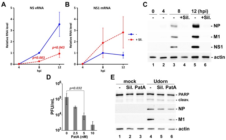
Eukaryotic translation initiation factor 4A (eIF4A) is a helicase that facilitates assembly of the translation preinitiation complex by unwinding structured mRNA 5' untranslated regions. Pateamine A (PatA) and silvestrol are natural products that disrupt eIF4A function and arrest translation, thereby triggering the formation of cytoplasmic aggregates of stalled preinitiation complexes known as stress granules (SGs). Here we examined the effects of eIF4A inhibition by PatA and silvestrol on influenza A virus (IAV) protein synthesis and replication in cell culture. Treatment of infected cells with either PatA or silvestrol at early times post-infection resulted in SG formation, arrest of viral protein synthesis and failure to replicate the viral genome. PatA, which irreversibly binds to eIF4A, sustained long-term blockade of IAV replication following drug withdrawal, and inhibited IAV replication at concentrations that had minimal cytotoxicity. By contrast, the antiviral effects of silvestrol were fully reversible; drug withdrawal caused rapid SG dissolution and resumption of viral protein synthesis. IAV inhibition by silvestrol was invariably associated with cytotoxicity. PatA blocked replication of genetically divergent IAV strains, suggesting common dependence on host eIF4A activity. This study demonstrates that the core host protein synthesis machinery can be targeted to block viral replication.

Keywords: eIF4A helicase; influenza A virus; pateamine A; silvestrol; stress granule; translation.


Conflict of interest statement

The authors declare no conflict of interest.

Figures

Figure 1
Figure 1
Concentration-dependent stress granule induction and inhibition of viral protein accumulation by pateamine A (PatA) and silvestrol (Sil.). A549, Vero, and MDCK cell lines infected with PR8 strain of influenza A virus (IAV) were treated with the indicated concentrations of PatA or Sil. at 1 hpi. (AC) Viral protein accumulation was analyzed in whole cell lysates collected at 24 hpi by western blot. (D) Stress granule induction and viral protein accumulation were visualized at 9 hpi by immunofluorescence microscopy using an antibody to stress granule marker TIAR (red) and a polyclonal anti-influenza antibody (green). Nuclei were stained with Hoechst dye (blue). Representative images are shown for each cell line and treatment condition. There were three biological replicates of A549 cell experiments, and two biological replicates for Vero and MDCK cell experiments.
Figure 2
Figure 2
Antiviral and cytotoxic effects of pateamine A and silvestrol vary between cell types. (A,B) Production of infectious virus progeny (PR8 strain) at 24 hpi was measured using plaque assay. The indicated cell lines were infected with multiplicity of infection (MOI) = 0.1 and treated with the increasing concentrations of pateamine A (A) or silvestrol (B) at 1 hpi. Dotted horizontal lines in A and B indicate the average PFU/mL of untreated cells. (C,D) Cell viability was measured using Alamar Blue assay after 24-h treatment with increasing concentrations of pateamine A (C) or silvestrol (D). Relative fluorescence values are normalized to vehicle control (DMSO). All error bars represent standard deviations from 3 independent biological replicates. Dotted horizontal lines in (C,D) indicate the relative fluorescence of untreated cells.
Figure 3
Figure 3
Translation inhibition by silvestrol is fully reversible. (A) A549 cells infected with PR8 strain of IAV were treated at 1 hpi with 320 nM silvestrol (Sil.) or 20 nM pateamine A (PatA) or left untreated. At 4 hpi, some wells were washed briefly with PBS and received fresh infection media without drugs as shown on the schematic outline of the experiment. At 12 hpi, mock and virus-infected cells subjected to continuous incubation with Sil. or PatA, or to drug wash off (WO) at 4 hpi, were analyzed by immunofluorescence staining with the polyclonal anti-influenza antibody (IAV, green) and the antibodies to SG markers TIAR (red) and G3BP (blue). (B) Total translation rates in A549 cells were analyzed using metabolic labelling with puromycin and subsequent western blotting with anti-puromycin antibody. In some cases, after initial three-hour treatments, Sil. or PatA were washed off prior to puromycin labeling. Total protein was visualized using BioRad Stain-Free reagent.
Figure 3
Figure 3
Translation inhibition by silvestrol is fully reversible. (A) A549 cells infected with PR8 strain of IAV were treated at 1 hpi with 320 nM silvestrol (Sil.) or 20 nM pateamine A (PatA) or left untreated. At 4 hpi, some wells were washed briefly with PBS and received fresh infection media without drugs as shown on the schematic outline of the experiment. At 12 hpi, mock and virus-infected cells subjected to continuous incubation with Sil. or PatA, or to drug wash off (WO) at 4 hpi, were analyzed by immunofluorescence staining with the polyclonal anti-influenza antibody (IAV, green) and the antibodies to SG markers TIAR (red) and G3BP (blue). (B) Total translation rates in A549 cells were analyzed using metabolic labelling with puromycin and subsequent western blotting with anti-puromycin antibody. In some cases, after initial three-hour treatments, Sil. or PatA were washed off prior to puromycin labeling. Total protein was visualized using BioRad Stain-Free reagent.
Figure 4
Figure 4
Sustained eIF4A inhibition by silvestrol and pateamine A leads to apoptosis. (A) Western blot analysis of A549 cell lysates obtained at the indicated times post-PR8-infection and treated with 320 nM silvestrol (Sil.) or 20 nM pateamine A (PatA) at 4 hpi or the equivalent time after mock infection. (B) Total RNA was isolated from cells treated with 320 nM silvestrol at 4 hpi and the relative levels of viral NS1 mRNA and vRNA at the indicated times post-infection were determined using RT-qPCR. Error bars represent standard deviations (n = 3). p values were calculated using a paired Student’s t-test. (C) Western blotting analysis of 16 hpi lysates of A549 wild type (WT) or PKR knock-out (PKR KO) cells mock infected or infected with PR8 strain of IAV. Cells were treated with 320 nM silvestrol (Sil.) or 20 nM pateamine A (PatA) at 4 hpi or the equivalent time after mock infection.
Figure 5
Figure 5
Silvestrol and pateamine A block replication of H3N2 strain of IAV. (AC) A549 cells were infected with A/Udorn/72(H3N2) strain of IAV and treated with 320 nM silvestrol at 4 hpi. Total RNA and whole cell protein lysates were collected at 4, 8, and 12 hpi. The accumulation of viral NS segment vRNA (A) and NS1 mRNA (B) was measured using RT-qPCR, and the accumulation of viral proteins was analysed by western blotting (C). In (A) and (B), error bars represent standard deviations (n = 3). (D) Production of infectious virus progeny (Udorn strain) at 24 hpi was measured using plaque assay. A549 cells were infected with MOI = 0.1 and treated with the increasing concentrations of pateamine A at 1 hpi. Error bars represent standard deviations (n = 4). (E) Western blotting analysis of A549 cell lysates obtained at 24 h post-infection with the Udorn strain of IAV and treated with 40 nM silvestrol (Sil.) or 5 nM pateamine A (PatA) at 4 hpi or the equivalent time after mock infection. p values in (A) and (D) were calculated using paired Student’s t-test.

References

    1. Plotch S.J., Bouloy M., Ulmanen I., Krug R.M. A unique cap(m7GpppXm)-dependent influenza virion endonuclease cleaves capped RNAs to generate the primers that initiate viral RNA transcription. Cell. 1981;23:847–858. doi: 10.1016/0092-8674(81)90449-9. - DOI - PubMed
    1. Khaperskyy D.A., Schmaling S., Larkins-Ford J., McCormick C., Gaglia M.M. Selective degradation of host RNA polymerase II transcripts by influenza A virus PA-X host shutoff protein. PLoS Pathog. 2016;12:e1005427. doi: 10.1371/journal.ppat.1005427. - DOI - PMC - PubMed
    1. Nemeroff M.E., Barabino S.M., Li Y., Keller W., Krug R.M. Influenza virus NS1 protein interacts with the cellular 30 kDa subunit of CPSF and inhibits 3’end formation of cellular pre-mRNAs. Mol. Cell. 1998;1:991–1000. doi: 10.1016/S1097-2765(00)80099-4. - DOI - PubMed
    1. Pereira C.F., Read E.K.C., Wise H.M., Amorim M.J., Digard P. Influenza A virus NS1 protein promotes efficient nuclear export of unspliced viral M1 mRNA. J. Virol. 2017;91:e00528-17. doi: 10.1128/JVI.00528-17. - DOI - PMC - PubMed
    1. Satterly N., Tsai P.L., van Deursen J., Nussenzveig D.R., Wang Y., Faria P.A., Levay A., Levy D.E., Fontoura B.M. Influenza virus targets the mRNA export machinery and the nuclear pore complex. Proc. Natl. Acad. Sci. USA. 2007;104:1853–1858. doi: 10.1073/pnas.0610977104. - DOI - PMC - PubMed

MeSH terms

LinkOut - more resources