

A case of proliferative diabetic retinopathy in which scintillating particles appeared in the intravitreal cavity after laser photocoagulation
- PMID: 29258460
- PMCID: PMC5735882
- DOI: 10.1186/s12886-017-0654-5
A case of proliferative diabetic retinopathy in which scintillating particles appeared in the intravitreal cavity after laser photocoagulation
Abstract
Background: To report a case of proliferative diabetic retinopathy (PDR) exhibiting the appearance of scintillating particles presumed to be crystallin inside the intravitreal cavity after laser photocoagulation.
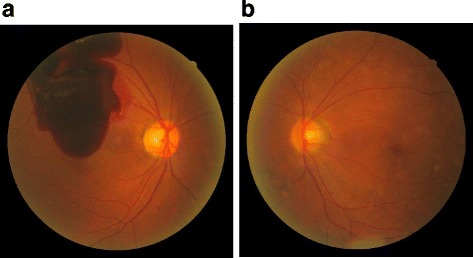
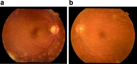
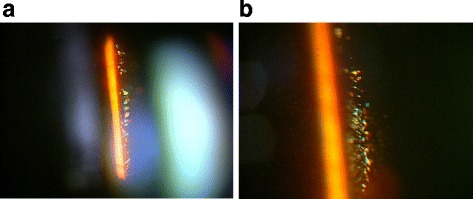
Case presentation: A 56-year-old male patient presented at our outpatient clinic after becoming aware of decreased vision in his right eye. Ocular examination performed at the patient's initial visit revealed a massive preretinal macular hemorrhage due to PDR in his right eye. Fundus fluorescein angiography revealed extensive retinal non-perfusion areas and neovascularization in both eyes. However, no opacity was observed in the intravitreal cavity of his left eye. Vitreous surgery was performed on the patient's right eye after ultrasonic phacoemulsification aspiration and intraocular lens implantation. Post surgery, the corrected VA in that eye improved from 0.1 to 1.0. In correlation with the treatment performed on the patient's right eye, we began panretinal photocoagulation on his left eye. Examination performed prior to the patient's third session of panretinal photocoagulation revealed a large number of scintillating particles in the posterior vitreous gel in front of the retina. Examination via slit-lamp microscopy revealed that the particles were of varied hues, and closely resembled a 'Christmas tree' cataract. No posterior vitreous detachment was observed, and since these particles were situated as if captured in the posterior vitreous gel, no eye-movement-associated mobility of the particles was observed. Since the cloudiness was not severe enough to interfere with photocoagulation, additional photocoagulation was performed, and the patient is currently under observation. Six months have now passed since the fourth photocoagulation procedure was performed, and there has been no change in the state of the particles. Optical coherence tomography imaging revealed no change before and after the panretinal photocoagulation. The corrected VA in his left eye has remained at 1.0 during the postoperative follow-up period.
Conclusions: We speculate that the production of crystallin in the retina in this case was triggered by the photocoagulation procedure performed for diabetic retinopathy.
Keywords: Crystallin; Laser photocoagulation; Posterior vitreous gel; Proliferative diabetic retinopathy; Scintillating particl.
Conflict of interest statement
Ethics approval and consent to participate
This case study was approved by the Ethics Committee of the Osaka Medical College.
Consent for publication
Written informed consent for publication was obtained from the patient.
Competing interests
The authors declare that they have no competing interests.
Publisher’s Note
Springer Nature remains neutral with regard to jurisdictional claims in published maps and institutional affiliations.
Figures

References
-
- Kase S, Ishida S, Rao NA. Increased expression of αA-crystallin in human diabetic eye. Int J Mol Med. 2011;28(4):505–511. - PubMed
Publication types
MeSH terms
Substances
LinkOut - more resources
Full Text Sources
Other Literature Sources
Medical
