

Anatomical variations of the palmaris longus muscle including its relation to the median nerve - a proposal for a new classification
- PMID: 29258498
- PMCID: PMC5738140
- DOI: 10.1186/s12891-017-1901-x
Anatomical variations of the palmaris longus muscle including its relation to the median nerve - a proposal for a new classification
Abstract
Background: The palmaris longus (PL) muscle is characterised by high morphological diversity, and its tendon crosses the median nerve (MN) at different levels. Due to the fact that the palmaris longus tendon is routinely harvested for reconstruction of other tendons, knowledge of its morphological variations is clinically important. Therefore, the purpose of the study was to suggest a new morphological classification of the PL muscle and characterise the relationship of its tendon to the median nerve.
Methods: Standard dissection was performed on 80 randomised and isolated upper limbs (40 left and 40 right) fixed in a 10% formalin solution. Measurements of muscle belly and tendon were obtained. The course and location of tendon insertion, as well as its relationship to the median nerve, were noted.
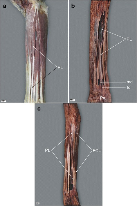
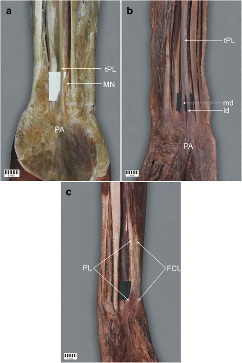
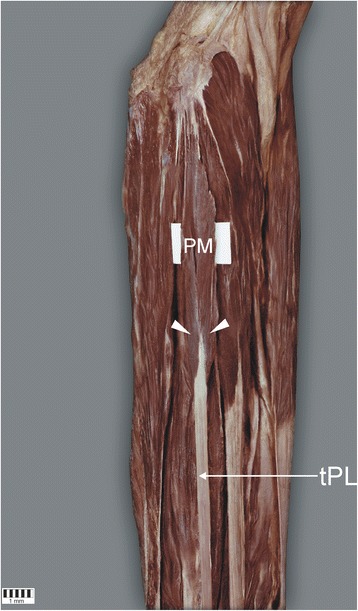
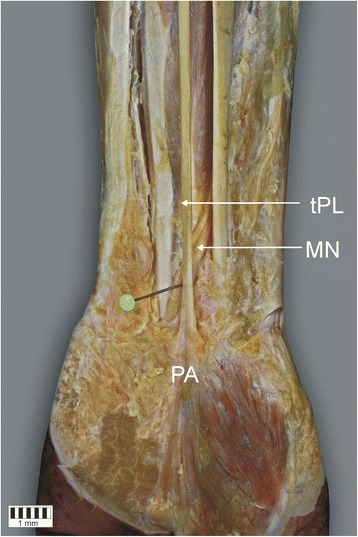
Results: The palmaris longus muscle was present in 92.5% of specimens. Three types of palmaris longus muscle were identified based on the morphology of its insertion (types I-III) and these were further subdivided into three subgroups (A-C) according to the ratio of the length of the muscle belly and its tendon. The most frequent was type I (78.8%), where the tendon attached to the palmar aponeurosis, and subtype B, where the tendon-to-belly ratio was 1-1.5 (41.1%). The mean distance from the interstyloid line to the crossing between the median nerve and the palmaris longus tendon was 31.6 mm. In addition, two types of palmaris longus were described.
Conclusion: The presented classification of palmaris longus muscle types allows a better characterization of its diversity and may be useful in planning tendon grafting.
Keywords: New classification; Palmaris longus muscle; Palmaris longus tendon; Tendon grafts.
Conflict of interest statement
Ethics approval and consent to participate
The protocol of the study was accepted by Bioethics Committee of Medical University of Lodz (resolution RNN/92/16/KE). The cadavers belong to the Department of Normal and Clinical Anatomy of the Medical University of Lodz. Each of the persons who died prematurely signed a notarial consent for the use of corpses for scientific and research purposes.
Consent for publication
Not applicable
Competing interests
The authors declare that they have no competing interests.
Publisher’s Note
Springer Nature remains neutral with regard to jurisdictional claims in published maps and institutional affiliations.
Figures
Similar articles
-
Morphological variability of the palmaris longus muscle in human fetuses.Surg Radiol Anat. 2018 Nov;40(11):1283-1291. doi: 10.1007/s00276-018-2069-2. Epub 2018 Jul 18. Surg Radiol Anat. 2018. PMID: 30022223 Free PMC article.
-
The palmar cutaneous branch of the median nerve and the palmaris longus tendon: a cadaveric study.J Hand Surg Am. 1994 Mar;19(2):199-202. doi: 10.1016/0363-5023(94)90005-1. J Hand Surg Am. 1994. PMID: 8201180
-
Variation in the insertion of the palmaris longus tendon.Singapore Med J. 2015 Jan;56(1):e7-9. doi: 10.11622/smedj.2015018. Singapore Med J. 2015. PMID: 25640108 Free PMC article.
-
Bilateral reversed palmaris longus muscle: a case report and systematic literature review.Surg Radiol Anat. 2020 Mar;42(3):289-295. doi: 10.1007/s00276-019-02363-z. Epub 2019 Nov 12. Surg Radiol Anat. 2020. PMID: 31720753 Free PMC article.
-
Morphometric analysis and surgical adequacy of palmaris longus as a tendon graft. A systematic review of cadaveric studies.Surg Radiol Anat. 2020 Mar;42(3):259-267. doi: 10.1007/s00276-019-02381-x. Epub 2019 Nov 18. Surg Radiol Anat. 2020. PMID: 31741040
Cited by
-
Nuances in endoscopic carpal tunnel release a guide to improving outcomes.J Hand Microsurg. 2024 Aug 22;16(5):100150. doi: 10.1016/j.jham.2024.100150. eCollection 2024 Dec. J Hand Microsurg. 2024. PMID: 39669728 Free PMC article. Review.
-
Is the plantaris muscle the most undefined human skeletal muscle?Anat Sci Int. 2021 Jun;96(3):471-477. doi: 10.1007/s12565-020-00586-4. Epub 2020 Nov 7. Anat Sci Int. 2021. PMID: 33159667 Free PMC article.
-
Morphological variability of the palmaris longus muscle in human fetuses.Surg Radiol Anat. 2018 Nov;40(11):1283-1291. doi: 10.1007/s00276-018-2069-2. Epub 2018 Jul 18. Surg Radiol Anat. 2018. PMID: 30022223 Free PMC article.
-
A proposal for a new classification of coracobrachialis muscle morphology.Surg Radiol Anat. 2021 May;43(5):679-688. doi: 10.1007/s00276-021-02700-1. Epub 2021 Feb 9. Surg Radiol Anat. 2021. PMID: 33564931 Free PMC article.
-
Anatomical Variants of the Upper Limb Nerves: Clinical and Preoperative Relevance.Semin Musculoskelet Radiol. 2023 Apr;27(2):129-135. doi: 10.1055/s-0043-1761952. Epub 2023 Apr 3. Semin Musculoskelet Radiol. 2023. PMID: 37011614 Free PMC article. Review.
References
-
- Mathew AJ. Versatile but Temperamental: A Morphological Study of Palmaris Longus in the Cadaver. J Clin Diagn Res. 2015; Available from: http://www.jcdr.net/article_fulltext.asp?issn=0973-709x&year=2015&volume... - PMC - PubMed
-
- Bergman RA, Afifi AK, Miyauchi R. Anatomical Variation, Radiology Anatomy, Anatomy Atlas. 2015. Anatomy Atlases: Illustrated Encyclopedia of Human Anatomic Variation.
MeSH terms
LinkOut - more resources
Full Text Sources
Other Literature Sources
