. 2017 Dec 19;14(1):252.
doi: 10.1186/s12974-017-1004-5.

Dendrimer-mediated delivery of N-acetyl cysteine to microglia in a mouse model of Rett syndrome

Affiliations

Dendrimer-mediated delivery of N-acetyl cysteine to microglia in a mouse model of Rett syndrome

Elizabeth Nance et al. J Neuroinflammation. .

Erratum in

Abstract

Background: Rett syndrome (RTT) is a pervasive developmental disorder that is progressive and has no effective cure. Immune dysregulation, oxidative stress, and excess glutamate in the brain mediated by glial dysfunction have been implicated in the pathogenesis and worsening of symptoms of RTT. In this study, we investigated a new nanotherapeutic approach to target glia for attenuation of brain inflammation/injury both in vitro and in vivo using a Mecp2-null mouse model of Rett syndrome.

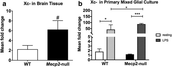
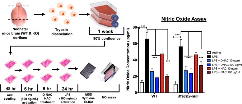
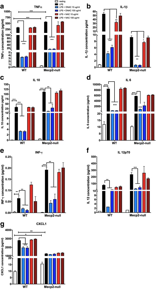
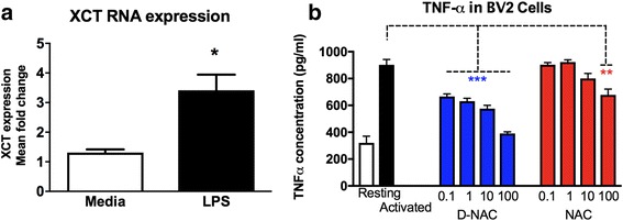
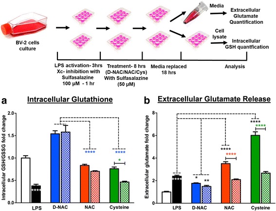
Methods: To determine whether inflammation and immune dysregulation were potential targets for dendrimer-based therapeutics in RTT, we assessed the immune response of primary glial cells from Mecp2-null and wild-type (WT) mice to LPS. Using dendrimers that intrinsically target activated microglia and astrocytes, we studied N-acetyl cysteine (NAC) and dendrimer-conjugated N-acetyl cysteine (D-NAC) effects on inflammatory cytokines by PCR and multiplex assay in WT vs Mecp2-null glia. Since the cysteine-glutamate antiporter (Xc-) is upregulated in Mecp2-null glia when compared to WT, the role of Xc- in the uptake of NAC and L-cysteine into the cell was compared to that of D-NAC using BV2 cells in vitro. We then assessed the ability of D-NAC given systemically twice weekly to Mecp2-null mice to improve behavioral phenotype and lifespan.

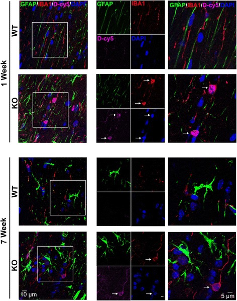
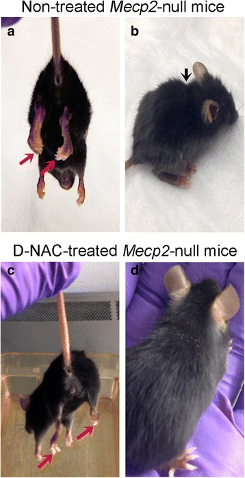
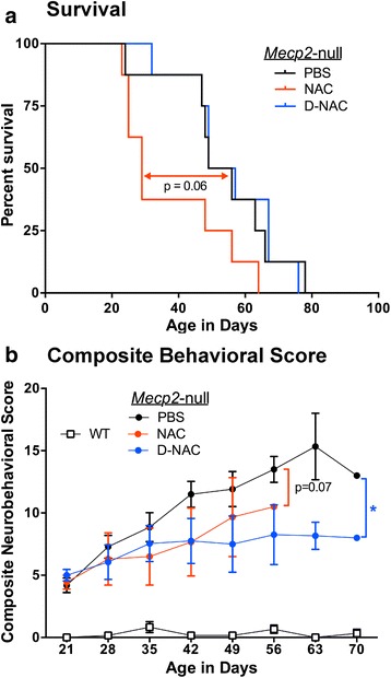
Results: We demonstrated that the mixed glia derived from Mecp2-null mice have an exaggerated inflammatory and oxidative stress response to LPS stimulation when compared to WT glia. Expression of Xc- was significantly upregulated in the Mecp2-null glia when compared to WT and was further increased in the presence of LPS stimulation. Unlike NAC, D-NAC bypasses the Xc- for cell uptake, increasing intracellular GSH levels while preventing extracellular glutamate release and excitotoxicity. Systemically administered dendrimers were localized in microglia in Mecp2-null mice, but not in age-matched WT littermates. Treatment with D-NAC significantly improved behavioral outcomes in Mecp2-null mice, but not survival.

Conclusions: These results suggest that delivery of drugs using dendrimer nanodevices offers a potential strategy for targeting glia and modulating oxidative stress and immune responses in RTT.

Keywords: Glutamate; Mecp2-null; Microglia; N-Acetyl cysteine; PAMAM dendrimer; Rett syndrome; System Xc−.


Conflict of interest statement

Ethics approval

Animal experiments were conducted within the guidelines set forth by the National Institutes of Health (NIH) and the Association for Assessment and Accreditation of Laboratory Animal Care (AAALAC) International. All animal protocols were approved by the Johns Hopkins Animal Care and Use Committee.

Consent for publication

Not applicable.

Competing interests

SK and RK hold a patent on dendrimer-based nanodevices for therapeutic and imaging purposes (US8889101) and are founders of a company focused on clinical translation of this compound.

Publisher’s Note

Springer Nature remains neutral with regard to jurisdictional claims in published maps and institutional affiliations.

Figures

Fig. 1
Fig. 1
XCT expression in wild-type (WT) versus Mecp2-null mice. a At 1 week of age, XCT mRNA expression was increased in Mecp2-null mice. b XCT mRNA expression increased significantly after stimulation with lipopolysaccharide toxin (LPS; checkered bars) in primary mixed glial cultures from WT and Mecp2-null mice. The response to LPS was also significantly increased in Mecp2-null compared to WT primary mixed glial cells. Mean values ± SEM for graphs in a and b (# p = 0.06; *p < 0.05; ***p < 0.001)
Fig. 2
Fig. 2
Increased nitric oxide (NO) release from Mecp2-null primary mixed glial cells challenged with LPS. Cortical tissue samples from the brains of 1-week-old Mecp2-null and WT mice were taken, and cells were dissociated and plated to grow. Once confluent, cells were plated, activated with LPS, and treated with D-NAC and NAC (left panel). Both WT and Mecp2-null glial cells showed equivalent NO release in response to LPS (black bars) that was diminished significantly by D-NAC (blue bars) administration (right panel). Only the high dose of free NAC was effective in decreasing the response to LPS (red bars)
Fig. 3
Fig. 3
Increased cytokine release in WT and Mecp2-null primary mixed glial cell cells challenged with LPS. A multiplex ELISA was conducted on the media collected from the mixed glial cell cultures. At rest (white bars), TNF-α (a), IL-10 (c), and CXCL1 (g) release was increased in Mecp2-null mixed glial culture compared to WT whereas release of IL-1β (b) was decreased. No group differences in IL-6, INF-γ, and IL-12 were observed at rest (df). After LPS stimulation (black bars), concentrations of TNF-α (a), IL-10 (c), INF-γ (e), and IL-12 (f) were significantly higher in Mecp2-null compared to WT glial cells. In contrast, blunting of the response to LPS stimulation for IL-1β (b), IL-6 (d), and CXCL1 (g) was observed in Mecp2-null glial cells compared to WT. Across the board, D-NAC 10 μg/mL and D-NAC 100 μg/mL (blue bars) consistently lowered all cytokine levels significantly in WT and Mecp2-null LPS-treated cells (all p ≤ 0.001) with the exception of the chemokine CXCL1 in Mecp2-null glial cells. In most cases, free NAC (red bars) was not effective except for IL-1β in which 10 μg/mL NAC decreased IL-1b levels significantly (*p < 0.05; **p < 0.01; ***p < 0.001)
Fig. 4
Fig. 4
Effects of LPS stimulation on XCT and TNF-α expression. a XCT expression in BV2 cells was increased after LPS stimulation (*p < 0.05). b BV2 cell cultures treated with D-NAC showed dose-dependent decreases in TNF-α concentration in the supernatant as compared to LPS-treated (activated) cultures. Only the highest dose of NAC (100 μg/mL) was effective at reducing TNF-α concentration
Fig. 5
Fig. 5
Role of Xc in NAC internalization in BV2 cells. Mecp2-null and WT brains were acquired, and cells were dissociated and plated to grow. Once confluent, cells were reseeded and treated with LPS (3 h) and sulfasalazine (1 h) and then incubated in cysteine, NAC, or D-NAC with sulfasalazine for 8 h. Then, the cells were incubated in fresh media for 18 h. Cells and media were then taken and used to assess intracellular glutathione levels and extracellular glutamate release. To assess the mechanism of internalization of D-NAC as compared to NAC and cysteine, extracellular glutamate and intracellular glutathione levels were assessed in LPS-activated BV2 cells after treatment when Xc was functional (without sulfasalazine (solid bars)) and when it was blocked with an Xc inhibitor (with sulfasalazine (cross-hatched bars)). a In D-NAC-treated cells, GSH increased regardless of Xc functionality/blockade (blue bars), suggesting that D-NAC bypasses Xc to exert its anti-oxidant effect intracellularly. In NAC- and cysteine-treated cells, Xc blockade resulted in a decrease in GSH production, suggesting that NAC and cysteine were internalized in large part via Xc. b Glutamate levels increased with LPS administration. When Xc was functional, NAC (red bars) and cysteine (green bars) showed increased glutamate levels, but when Xc was blocked, glutamate release decreased in NAC- and cysteine-treated samples. In contrast, regardless of Xc functionality/blockade, D-NAC (blue bars) was effective in reducing glutamate release (*p < 0.05; **p < 0.01; ***p < 0.001; white asterisks denote the comparison between resting and LPS conditions; the black asterisks denote the comparison between LPS-stimulated and LPS + D-NC, NAC, or cysteine conditions; the blue asterisks denote the comparison between LPS + D-NAC and LPS + NAC and LPS + cysteine conditions; the red and green asterisks denote the comparison between LPS + NAC and LPS + NAC + sulfasalazine and LPS + cysteine and LPS + cysteine + sulfasalazine)
Fig. 6
Fig. 6
Brain uptake and cellular localization of D-Cy5 in WT and Mecp2-null mice. Dendrimer labeled with Cy5 (D-Cy5; magenta) was taken into microglia (Iba1, red) in the pre-symptomatic period (1 week of age) and in the symptomatic period (7 weeks of age). Little-to-no uptake of D-Cy5 was observed in astrocytes (GFAP; green). White arrows indicated D-Cy5 co-localization with Iba+ cells. Cell nuclei were stained with DAPI (blue). Scale bars = 10 or 5 μm
Fig. 7
Fig. 7
The physical appearance of Mecp2-null mice was improved by D-NAC therapy. Non-treated Mecp2-null mice were emaciated and had severe paw clenching, hunched posture, and poor eye conditions (a, b). D-NAC improved the overall appearance of Mecp2-null mice (c, d)
Fig. 8
Fig. 8
Effects of D-NAC on neurobehavioral outcomes and survival in Mecp2-null mice. a Twice weekly injections of 10 mg/kg doses of D-NAC treatment significantly improved the composite behavioral score compared to vehicle (PBS)-injected Mecp2-null mice (*p < 0.05). There was a trend for the composite behavioral score to be lower in NAC-treated Mecp2-null mice (p = 0.07). b Survival was assessed using a Kaplan-Meier curve. D-NAC did not improve survival compared to PBS-treated animals. D-NAC-treated Mecp2-null mice showed a trend for longer survival time (p = 0.06) compared to NAC-treated mice

Similar articles

Cited by

References

    1. Lyst MJ, Bird A. Rett syndrome: a complex disorder with simple roots. Nat Rev Genet. 2015;16:1–13. doi: 10.1038/nrg3897. - DOI - PubMed
    1. Hagberg B, Goutières F, Hanefeld F, Rett A, Wilson J. Rett syndrome: criteria for inclusion and exclusion. Brain and Development. 1985;7:372–373. doi: 10.1016/S0387-7604(85)80048-6. - DOI - PubMed
    1. Naidu S, Murphy M, Moser HW, Rett A. Rett syndrome: natural history in 70 cases. Am J Med Genet. 1986;24:61–72. doi: 10.1002/ajmg.1320250507. - DOI - PubMed
    1. Rett A. History and general overview. Am J Med Genet. 1986;24:21–25. doi: 10.1002/ajmg.1320250503. - DOI - PubMed
    1. Neul JL, Kaufmann WE, Glaze DG, Christodoulou J, Clarke AJ, Bahi-Buisson N, et al. Rett syndrome: revised diagnostic criteria and nomenclature. Ann Neurol. 2010;68:944–950. doi: 10.1002/ana.22124. - DOI - PMC - PubMed

MeSH terms