. 2017 Dec 19;14(1):56.
doi: 10.1186/s12977-017-0382-1.

Moloney leukemia virus 10 (MOV10) inhibits the degradation of APOBEC3G through interference with the Vif-mediated ubiquitin-proteasome pathway

Affiliations

Moloney leukemia virus 10 (MOV10) inhibits the degradation of APOBEC3G through interference with the Vif-mediated ubiquitin-proteasome pathway

Cancan Chen et al. Retrovirology. .

Abstract

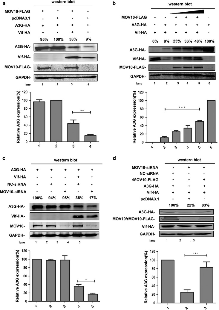
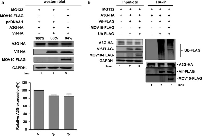
Background: MOV10 protein has ATP-dependent 5'-3' RNA helicase activity and belongs to the UPF1p superfamily. It can inhibit human immunodeficiency virus type 1 (HIV-1) replication at multiple stages and interact with apolipoprotein-B-mRNA-editing enzyme catalytic polypeptide-like 3G (APOBEC3G or A3G), a member of the cytidine deaminase family that exerts potent inhibitory effects against HIV-1 infection. However, HIV-1-encoded virion infectivity factor (Vif) protein specifically mediates the degradation of A3G via the ubiquitin-proteasome system (UPS).

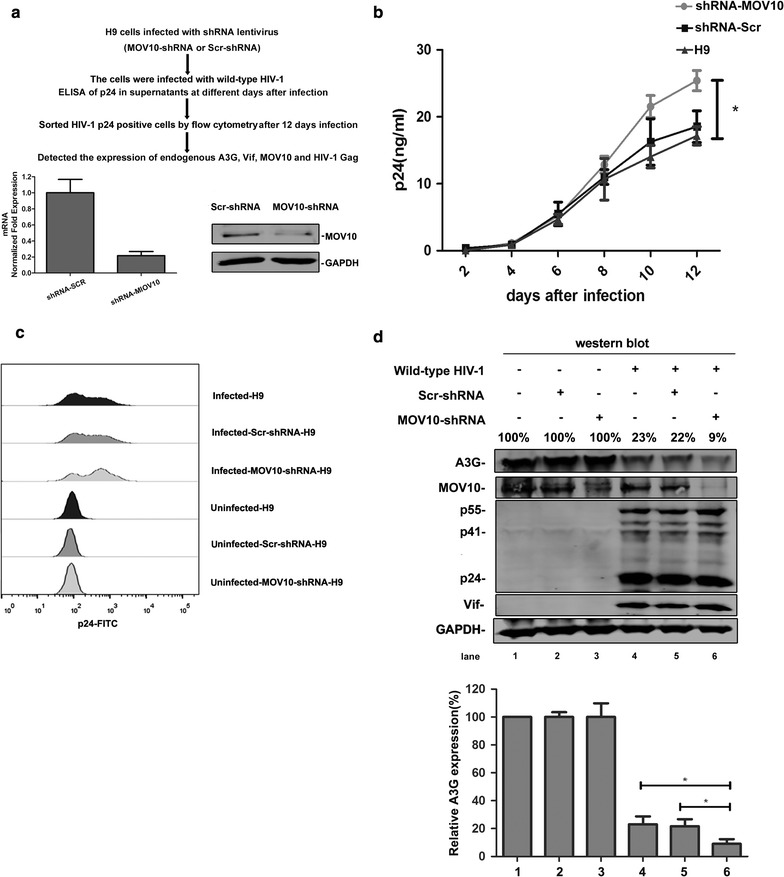
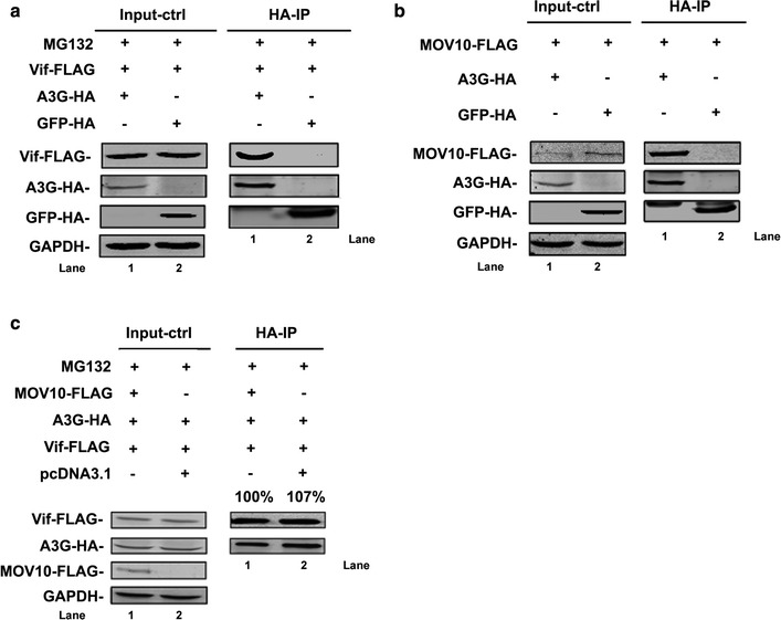
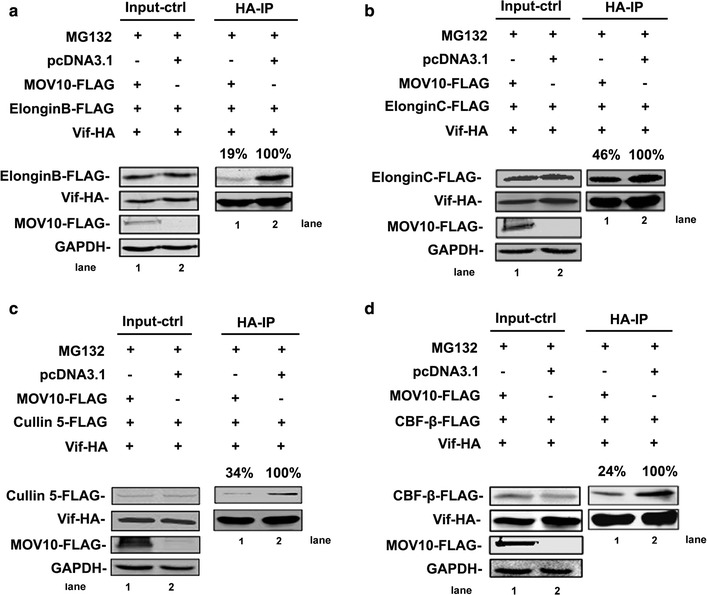
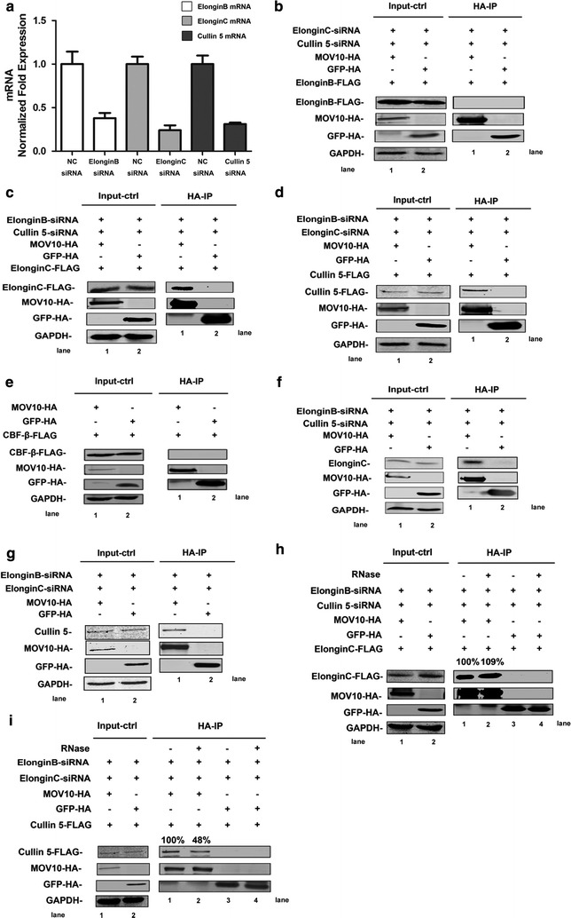
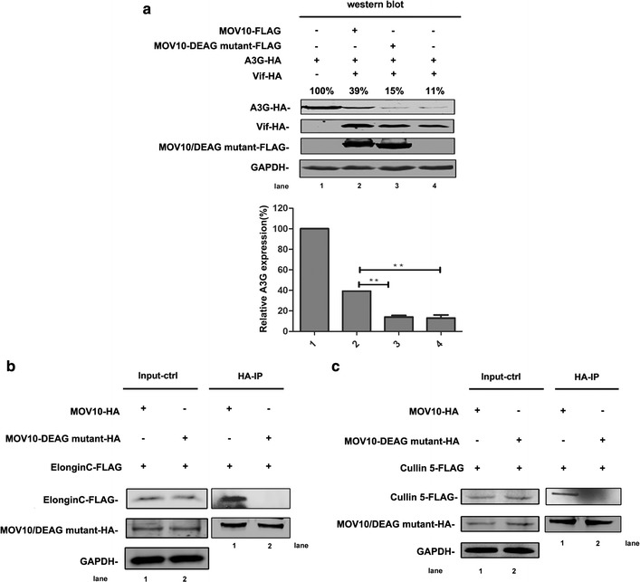
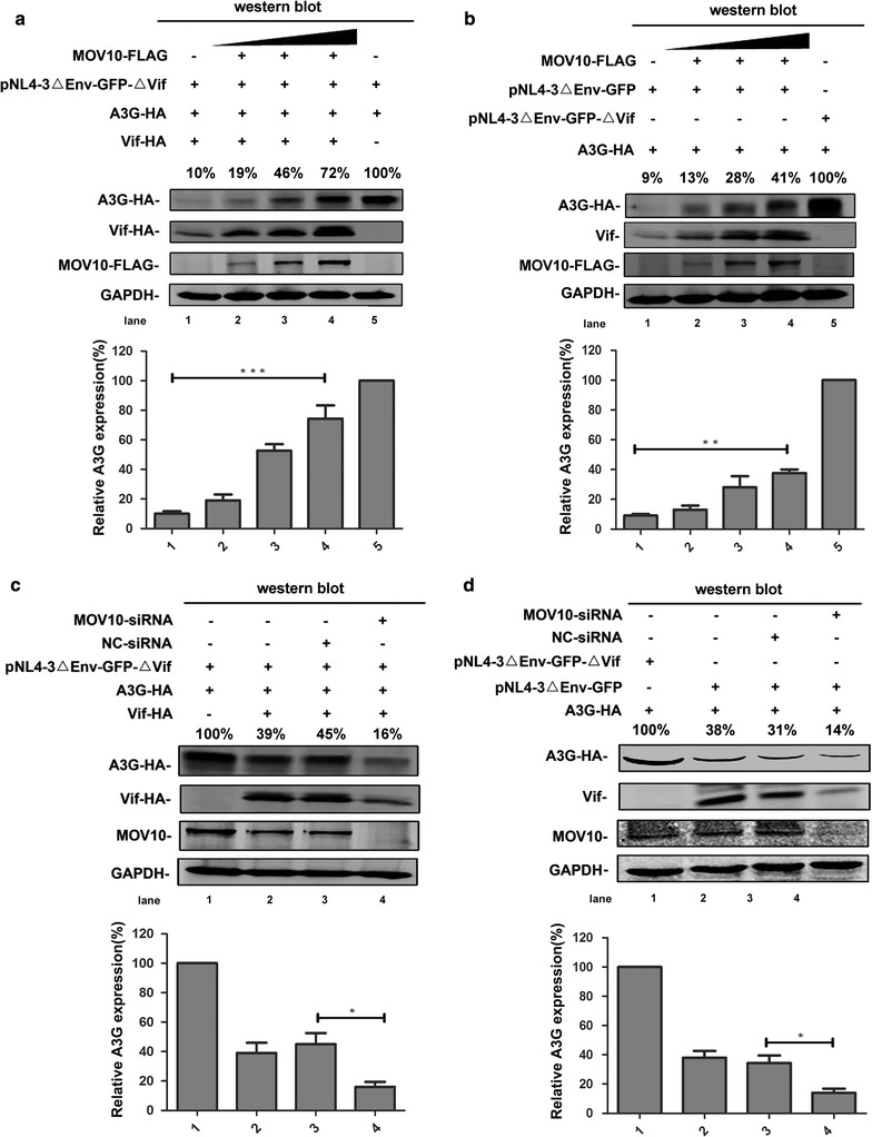
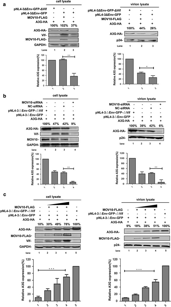
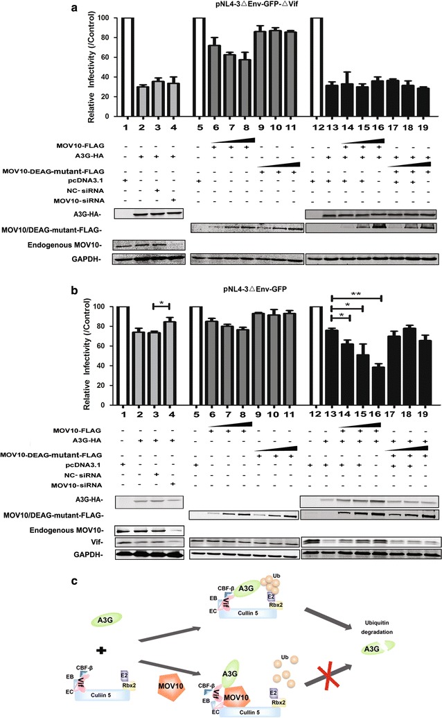
Results: We demonstrate that MOV10 counteracts Vif-mediated degradation of A3G by inhibiting the assembly of the Vif-CBF-β-Cullin 5-ElonginB-ElonginC complex. Through interference with UPS, MOV10 enhances the level of A3G in HIV-1-infected cells and virions, and synergistically inhibits the replication and infectivity of HIV-1. In addition, the DEAG-box of MOV10 is required for inhibition of Vif-mediated A3G degradation as the DEAG-box mutant significantly loses this ability.

Conclusions: Our results demonstrate a novel mechanism involved in the anti-HIV-1 function of MOV10. Given that both MOV10 and A3G belong to the interferon antiviral system, their synergistic inhibition of HIV-1 suggests that these proteins may play complicated roles in antiviral functions.

Keywords: A3G; HIV-1; MOV10; Ubiquitin–proteasome system (UPS); Vif.


Figures

Fig. 1
Fig. 1
MOV10 counteracts Vif-mediated degradation of A3G. a, b MOV10 overexpression inhibits Vif-mediated A3G degradation. Human 293T cells were transfected with pcDNA3.1-A3G-HA (0.8 μg), pcDNA3.1-Vif-HA (0.5 μg), and pcDNA3.1-MOV10-FLAG (1.5 μg) (a) or different amounts of pcDNA3.1-MOV10-FLAG (from 0.5 to 2 μg) (b). Then, cells were collected and lysed at 48 h, and analyzed by western blotting with anti-HA, anti-FLAG, and anti-GAPDH antibodies. c, d The effect of MOV10 depletion on Vif-mediated degradation of A3G. Cells were transfected with pcDNA3.1-A3G-HA (0.8 μg), pcDNA3.1-Vif-HA (0.5 μg), MOV10-specific siRNA (50 nM) (c) and/or siRNA-resistant MOV10 construct (rMOV10-FLAG) (0.4 μg) (d). After 48 h, cells were collected and analyzed by western blotting assay with anti-HA, anti-FLAG, anti-MOV10 and anti-GAPDH antibodies. Empty vector pcDNA3.1 was used in each transfection to normalize DNA amounts. Values in ad represent percentages of A3G or MOV10 normalized against GAPDH and compared with control. The bar graphs represent the average expression of A3G with different treatment and relative to the A3G-only reaction control (set to 100%). All the data represent mean ± SD from three independent experiments. Statistical significance was determined using t test: *p ≤ 0.05; **p ≤ 0.01; ***p ≤ 0.001
Fig. 2
Fig. 2
MOV10 protects A3G from Vif-mediated degradation in wild-type HIV-1. a H9 cells were infected with pLKO.1-MOV10-shRNA or pLKO.1-Scr-shRNA lentivirus for 8 h and then selected with puromycin for 2 weeks. MOV10-knockdown H9 cells and control cells were infected with wild-type HIV-1 for 3 h and cultured with fresh medium for 12 days. The culture supernatants were collected at the indicated time points. Then HIV-1 p24 was detected using HIV-1 p24 ELISA kit at different time points (b). And at 12th day, these cells were analyzed by flow cytometer (c). HIV-1 p24 positive H9 cells were sorted and detected by western blotting with anti-MOV10, anti-A3G, anti-Vif, anti-HIV-1 p24, and anti-GAPDH antibodies (d). Values in d represent percentages of A3G or MOV10 normalized against GAPDH and compared with control. The bar graphs in d represent the average expression of A3G with different treatment and relative to the A3G-only reaction control (set to 100%). Data in a, b, and d represent mean ± SD from three independent experiments. *, statistically significant, p ≤ 0.05 (t test). All the results are representative of at least three independent experiments
Fig. 3
Fig. 3
MOV10 prevents A3G from Vif-induced degradation by decreasing the ubiquitination of A3G. a Human 293T cells were transfected with pcDNA3.1-A3G-HA (0.8 μg), pcDNA3.1-Vif-HA (0.5 μg), and pcDNA3.1-MOV10-FLAG (1.5 μg) and then treated with MG132 (4 μM) for 16 h. Lysed cells were collected at 48 h and detected by western blotting with anti-HA, anti-FLAG, and anti-GAPDH antibodies. b 293T cells were transfected with pcDNA3.1-A3G-HA (2 μg), pcDNA3.1-Vif-FLAG (1.25 μg), pcDNA3.1-MOV10-FLAG (2.5 μg), and pcDNA3.1-Ub-FLAG (3 μg). Cells were treated with MG132 (4 μM) for 16 h and analyzed by co-immunoprecipitation with anti-HA agarose beads. And then, samples were detected by western-blotting using anti-HA, anti-FLAG, and anti-GAPDH. Values in a represent percentages of A3G normalized against GAPDH and compared with control. The bar graphs in a represent the average expression of A3G with different treatment and relative to the A3G-only reaction control (set to 100%). Data in a represent mean ± SD from three independent experiments. Empty vector pcDNA3.1 was used to equalize DNA amounts in each transfection. Data in a and b are representative of at least three independent experiments
Fig. 4
Fig. 4
MOV10 has no influence on the binding of A3G with Vif. a, b A3G interacts with Vif or MOV10 effectively. a Human 293T cells were transfected with 2 μg of pcDNA3.1-A3G-HA (pcDNA3.1-GFP-HA as a control) and 1 μg of pcDNA3.1-Vif-FLAG and then treated with MG132 for 16 h. b Human 293T cells were transfected with 1 μg of pcDNA3.1-Vif-FLAG and 2 μg of pcDNA3.1-A3G-HA or pcDNA3.1-GFP-HA. a, b lysates from these transfected cell samples were subjected to co-immunoprecipitation analysis using anti-HA agarose beads and then detected by western blotting. c The effect of MOV10 on the interaction between A3G and Vif. 293T cells were transfected with 2 μg of pcDNA3.1-A3G-HA together with 1 μg of pcDNA3.1-Vif-FLAG, and 2 μg of pcDNA3.1-MOV10-FLAG and then treated with MG132 (4 μM) for 16 h. Samples were immunoprecipitated with anti-HA agarose beads and analyzed by western blotting. Empty vector pcDNA3.1 was used to equalize DNA amounts in each transfection. Values in c represent portions of Vif-FLAG normalized against A3G-HA relative to control values. Data in a, b, and c are representative of at least three independent experiments
Fig. 5
Fig. 5
MOV10 affects the assembly of Vif-CBF-β-Cullin 5-ElonginB-ElonginC Complex. ad The effect of MOV10 on the interaction between Vif and ElonginB (a), ElonginC (b), Cullin 5 (c), or CBF-β (d). 293T cells were transfected with pcDNA3.1-MOV10-FLAG (2 μg), pcDNA3.1-Vif-HA (1 μg), and 4 μg of ElonginB-FLAG (a) or ElonginC-FLAG (b) or pcDNA3.1-Cullin 5-FLAG (c) or pcDNA3.1-CBF-β-FLAG (d). After treated with MG132 (4 μM) for 16 h, cell lysates were immunoprecipitated with anti-HA agarose beads and analyzed by immunoblotting using anti-FLAG, anti-HA, and anti-GAPDH antibodies. In each transfection, empty vector pcDNA3.1 was used to normalize DNA amounts. Values in ad represent percentages of ElonginB-FLAG/ElonginC-FLAG/Cullin 5-FLAG/CBF-β-FLAG normalized against Vif-HA relative to control values. All the data is representative of at least three independent experiments
Fig. 6
Fig. 6
MOV10 binds with ElonginC or Cullin 5. a The knockdown efficiency of siElonginB, siElonginC and siCullin 5. 293T cells were transfected with siElonginB, siElonginC or siCullin 5, after 48 h, the cells were collected and detected with qRT-PCR. Data in A represents mean ± SD (error bars). bi Co-immunoprecipitated analysis of the interaction between MOV10 and ElonginB (b), ElonginC (c, f, h), Cullin 5 (d, g, i), or CBF-β (e). pcDNA3.1-ElonginB-FLAG plus siElonginC and siCullin 5 (b), pcDNA3.1-ElonginC-FLAG plus siElonginB and siCullin 5 (c, h), pcDNA3.1-Cullin 5-FLAG plus siElonginB and siEloingC (d, i) or pcDNA3.1-CBF-β-FLAG (e) was transfected into 293T cells with pcDNA3.1-MOV10-HA or pcDNA3.1-GFP-HA. 293T cells were transfected with pcDNA3.1-MOV10-HA (pcDNA3.1-GFP-HA as a control) plus siElonginB and siCullin 5 (f) or siElonginB and siEloingC (g). After 48 h, the cells were collected and immunoprecipitated with anti-HA agarose beads (bi). The samples in h and i were treated with RNase mixture. And then, immunoprecipitated samples were analyzed by immunoblotting with anti-FLAG, anti-HA, anti-GAPDH, anti-MOV10, anti-ElonginC, and anti-Cullin 5 antibodies. Empty vector pcDNA3.1 was used to equalize DNA amounts in each transfection. Values in h and i represent portions of ElonginC-FLAG/Cullin 5-FLAG normalized against MOV10-HA and compared with control. Data in ai is representative of at least three independent experiments
Fig. 7
Fig. 7
The DEAG-box motif of MOV10 is required for the binding of MOV10 with ElonginC or Cullin 5. a The effect of MOV10-DEAG mutant on Vif-mediated A3G degradation. 293T cells were transfected with 0.4 μg of pcDNA3.1-Vif-HA, 0.8 μg of pcDNA3.1-A3G-HA, and 1.5 μg of pcDNA3.1-MOV10-FLAG or pcDNA3.1-MOV10-DEAG-mutant-FLAG as indicated. After 48 h, cell lysates were detected by western blotting assay with anti-HA, anti-FLAG, and anti-GAPDH antibodies. Values represent portions of A3G-HA normalized against GAPDH and compared with control. b, c Co-immunoprecipitated analysis of the interaction between MOV10-DEAG mutant and ElonginC or Cullin 5. Human 293T cells were transfected with 2 μg of pcDNA3.1-MOV10-HA or pcDNA3.1-MOV10-DEAG-HA and 6 μg of pcDNA3.1-ElonginC-FLAG or pcDNA3.1-Cullin 5-FLAG. After 24 h, MG132 were added in the transfected cells for 16 h. Then, the cells were collected for co-immunoprecipitation analysis with anti-HA agarose beads and detected by western blotting with anti-HA, anti-FLAG, and anti-GAPDH antibodies. In each transfection, empty vector pcDNA3.1 was used to equalize DNA amounts. Values in a represent percentages of A3G-HA normalized against GAPDH relative to control. The bar graphs in a represent the average expression of A3G with different treatments and relative to the A3G-only reaction control (set to 100%). Data in a represent mean ± SD from three independent experiments. Statistical significance was determined using t test: **p ≤ 0.01. All the data in a, b, and c is representative of at least three independent experiments
Fig. 8
Fig. 8
MOV10 reduces A3G proteasomal degradation significantly in the context of HIV-1 replication. a, b Overexpression of MOV10 inhibits Vif-induced A3G degradation in the context of HIV-1 replication. Human 293T cells were transfected with 0.8 μg of pcDNA3.1-A3G-HA, different amounts of pcDNA3.1-MOV10-FLAG (from 0.5 μg to 2 μg), 0.5 μg of pcDNA3.1-Vif-HA, 1 μg of pNL4-3ΔEnv-GFP-ΔVif (a) and/or 1 μg of pNL4-3ΔEnv-GFP (b) as indicated. Then, cells were collected at 48 h for western blotting assay with anti-FLAG, anti-HA, anti-Vif, and anti-GAPDH antibodies. c, d The effect of MOV10 depletion on the proteasomal degradation of A3G in the context of other HIV-1 proteins. 293T cells were transfected with pcDNA3.1-A3G-HA (0.8 μg), pcDNA3.1-Vif-HA (0.5 μg), MOV10-specific siRNA (or negative control-siRNA), 1 μg of pNL4-3ΔEnv-GFP-ΔVif (c) and/or 1 μg of pNL4-3ΔEnv-GFP (d). Cell lysates were detected by immunoblotting with anti-HA, anti-FLAG, anti-MOV10, anti-Vif, and anti-GAPDH antibodies. The bar graphs represent the average expression of A3G with different treatment and relative to the A3G-only reaction control (set to 100%). Data in ad represent mean ± SD from three independent experiments. Statistical significance was determined using t test: *p ≤ 0.05; **p ≤ 0.01; ***p ≤ 0.001. Empty vector pcDNA3.1 was used to equalize DNA amounts in each transfection. Values in ad represent portions of A3G-HA normalized against GAPDH and compared with control. Each data is representative of at least three independent experiments
Fig. 9
Fig. 9
MOV10 increases the quantity of A3G in HIV-1 virions. a Overexpression of MOV10 increases the quantity of A3G in virions. Human 293T cells were transfected with 0.8 μg of pcDNA3.1-A3G-HA, 1.5 μg of pcDNA3.1-MOV10-FLAG, 1 μg of pNL4-3ΔEnv-GFP or pNL4-3ΔEnv-GFP-ΔVif as indicated. b The effect of endogenous MOV10 knockdown on Vif-induced A3G degradation in HIV-1 virions. Cells were transfected with 0.8 μg of pcDNA3.1-A3G-HA, 50 nM of MOV10-specific siRNA (or negative control siRNA), and 1 μg of pNL4-3ΔEnv-GFP or pNL4-3ΔEnv-GFP-ΔVif. c MOV10 can be packaged into HIV-1 virions and its packaging level increases with the survival level of A3G. 293T cells were transfected with 0.8 μg of pcDNA3.1-A3G-HA, different amounts of pcDNA3.1-MOV10-FLAG (from 0.5 to 1.5 μg), 1 μg of pNL4-3ΔEnv-GFP or pNL4-3ΔEnv-GFP-ΔVif. a, b and c After 48 h, cell pellets and supernatants were collected respectively. Cell pellets were lysed and subjected to immunoblotting with anti-HA, anti-FLAG, anti-MOV10, anti-Vif, and anti-GAPDH antibodies. VLPs were collected from filtered supernatants by ultracentrifugation. The pelleted VLPs were lysed and detected by western blotting with anti-HA, anti-FLAG, and anti-p24 antibodies. The bar graphs represent the average expression of A3G with different treatments and relative to the A3G-only reaction control (set to 100%). Data in ac represent mean ± SD from three independent experiments. Statistical significance was determined using t test: *p ≤ 0.05; **p ≤ 0.01; ***p ≤ 0.001. In each transfection, empty vector pcDNA3.1 was used to equalize DNA amounts. Values in ac represent percentages of A3G-HA normalized against GAPDH or p24 and compared with control. Results are representative of at least three independent experiments
Fig. 10
Fig. 10
MOV10 synergistically enhances the inhibitory effect of A3G on the infectivity of HIV-1. a, b MOV10-specific siRNA was transfected in 293T cells with pcDNA3.1-A3G-HA (0.8 μg), pCMV-VSV-G (2.5 μg), and pNL4-3ΔEnv-GFP-ΔVif (7.5 μg) or pNL4-3ΔEnv-GFP (7.5 μg). 293T cells were co-transfected with pCMV-VSV-G (2.5 μg), pNL4-3ΔEnv-GFP-ΔVif (7.5 μg) or pNL4-3ΔEnv-GFP (7.5 μg), and increasing amounts of pcDNA3.1-MOV10-FLAG (0.5–1.5 μg) or pcDNA3.1-MOV10-DEAG-mutant-FLAG (0.5–1.5 μg) in the presence or absence of pcDNA3.1-A3G-HA (0.8 μg). Culture supernatants containing 5 ng of p24 were used to infect TZM-bl cells and luciferase activity was determined at 72 h post infection. For viruses with different amounts of MOV10 but no A3G, and viruses with a fixed amount of A3G combined with different amounts of MOV10 or not, the data are plotted as relative infectivity, with the control virus (pcDNA3.1) set to 100%. Error bars represent standard errors from three independent experiments. Statistical significance was determined using t test: *p ≤ 0.05; **p ≤ 0.01. c A cartoon to show the interaction between MOV10, A3G, and Vif. In the absence of MOV10, the Vif-CBF-β-Cullin 5-ElonginB-ElonginC complex is stable and triggers the proteasomal degradation of A3G. In the presence of MOV10, the assembly of Vif-CBF-β-Cullin 5-ElonginB-ElonginC complex can be disturbed by MOV10, which leads to A3G escaping from Vif-induced proteasomal degradation

Similar articles

Cited by

References

    1. Zhang H, Yang B, Pomerantz RJ, Zhang C, Arunachalam SC, Gao L. The cytidine deaminase CEM15 induces hypermutation in newly synthesized HIV-1 DNA. Nature. 2003;424:94–98. doi: 10.1038/nature01707. - DOI - PMC - PubMed
    1. Mariani R, Chen D, Schrofelbauer B, Navarro F, Konig R, Bollman B, Munk C, Nymark-McMahon H, Landau NR. Species-specific exclusion of APOBEC3G from HIV-1 virions by Vif. Cell. 2003;114:21–31. doi: 10.1016/S0092-8674(03)00515-4. - DOI - PubMed
    1. Lecossier D, Bouchonnet F, Clavel F, Hance AJ. Hypermutation of HIV-1 DNA in the absence of the Vif protein. Science. 2003;300:1112. doi: 10.1126/science.1083338. - DOI - PubMed
    1. Mangeat B, Turelli P, Caron G, Friedli M, Perrin L, Trono D. Broad antiretroviral defence by human APOBEC3G through lethal editing of nascent reverse transcripts. Nature. 2003;424:99–103. doi: 10.1038/nature01709. - DOI - PubMed
    1. Bourara K, Liegler TJ, Grant RM. Target cell APOBEC3C can induce limited G-to-A mutation in HIV-1. PLoS Pathog. 2007;3:1477–1485. doi: 10.1371/journal.ppat.0030153. - DOI - PMC - PubMed

MeSH terms