Review
. 2018 Jan;19(1):18-28.
doi: 10.15252/embr.201745130. Epub 2017 Dec 19.

Circadian clocks: from stem cells to tissue homeostasis and regeneration

Affiliations
Review

Circadian clocks: from stem cells to tissue homeostasis and regeneration

Pieterjan Dierickx et al. EMBO Rep. 2018 Jan.

Abstract

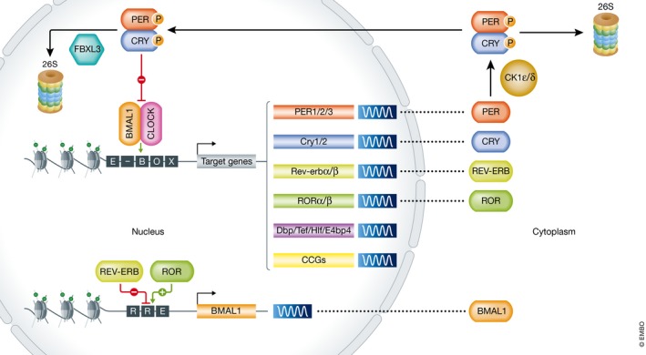
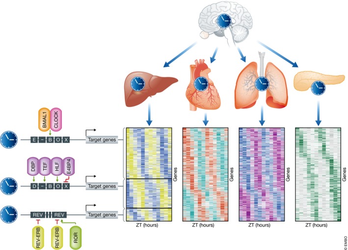
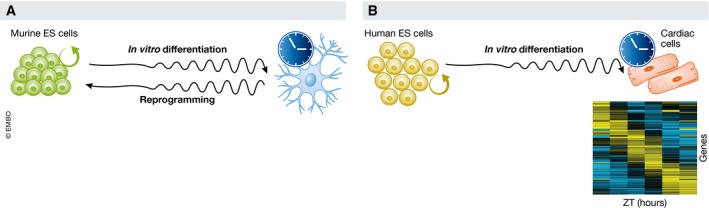
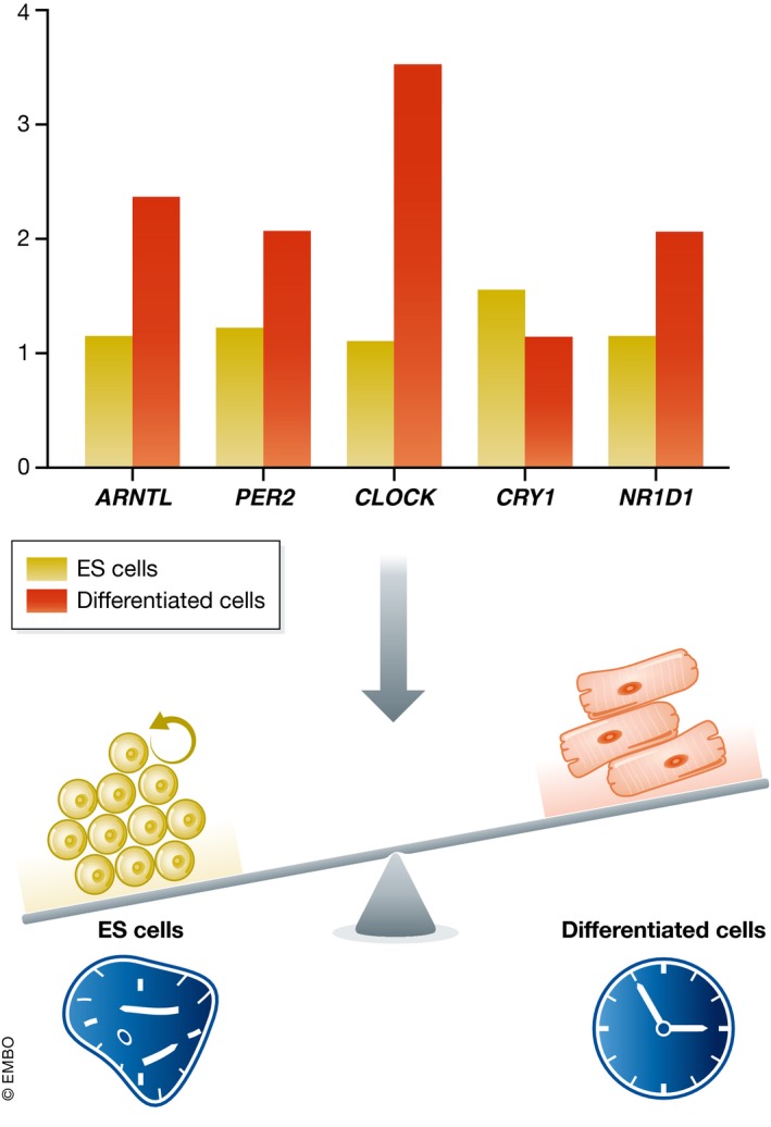
The circadian clock is an evolutionarily conserved timekeeper that adapts body physiology to diurnal cycles of around 24 h by influencing a wide variety of processes such as sleep-to-wake transitions, feeding and fasting patterns, body temperature, and hormone regulation. The molecular clock machinery comprises a pathway that is driven by rhythmic docking of the transcription factors BMAL1 and CLOCK on clock-controlled output genes, which results in tissue-specific oscillatory gene expression programs. Genetic as well as environmental perturbation of the circadian clock has been implicated in various diseases ranging from sleep to metabolic disorders and cancer development. Here, we review the origination of circadian rhythms in stem cells and their function in differentiated cells and organs. We describe how clocks influence stem cell maintenance and organ physiology, as well as how rhythmicity affects lineage commitment, tissue regeneration, and aging.

Keywords: aging; circadian rhythms; clock; regeneration; stem cells.


Figures

Figure 1
Figure 1. The molecular clock pathway
Schematic of the transcriptional/translational feedback loop. The BMAL1:CLOCK heterodimer binds enhancer (E)‐Box elements within regulatory sequences of its target genes, such as Per, Cry, Rev‐erb, Ror, Dbp, Tef, Hlf, E4bp4, and clock‐controlled genes (CCGs). Upon transcriptional induction of Per and Cry, PER and CRY proteins accumulate and dimerize in the cytosol, where they either get degraded by the 26S proteasome upon CK1ε/δ‐mediated phosphorylation or from where they migrate to the nucleus to inhibit BMAL1:CLOCK transcriptional activity and therefore their own transcription. Upon gradual phosphorylation and ubiquitination by FBXL3, they are degraded in the nucleus, completing the first feedback loop. The second BMAL1:CLOCK‐dependent feedback loop is driven by rhythmic Ror and Rev‐erb transcription. Upon accumulation of their respective proteins in the cytosol, ROR and REV‐ERB shuttle to the nucleus where they activate/repress Bmal1 transcription via competitive binding to the REV‐ERB/ROR response (RRE) element in its regulatory sequences. Additional post‐transcriptional/translational/epigenetic modifications mediate robustness of the pathway, thereby establishing cycles of around 24 h of rhythmic BMAL1:CLOCK‐mediated transcriptional activation of CCGs.
Figure 2
Figure 2. Organ‐specific clock‐controlled genes peak at different times during the circadian cycle
The central clock, located in the suprachiasmatic nucleus in the brain, synchronizes the clocks of peripheral clocks, which on their turn drive rhythmic expression of clock‐controlled genes (CCGs) that are often tissue‐specific (depicted as differentially colored heatmaps). This is mediated by tissue‐specific transcription factors that bind regulatory elements of CCGs, which results in peaks/phases of transcription at different ZTs (zeitgeber times).
Figure 3
Figure 3. The circadian clock during in vitro (de)differentiation
(A) Random differentiation of mouse embryonic stem (ES) cells leads to gradual activation of the molecular circadian clock, while reprogramming decreases rhythmicity of the expression of clock genes. (B) Directed differentiation of human ES cells toward the cardiac lineage leads to activation of the circadian clock that drives oscillatory gene expression of a set of clock‐controlled genes.
Figure 4
Figure 4. Altered clock factor stoichiometry might shift the functional role of core clock genes toward modulation of proliferation
ES cells express core clock factors (ARNTL, PER2, CLOCK, CRY1, and NR1D1) at a different level than differentiated cells. These factors may exert non‐circadian roles in proliferative ES cells that is distinct from their circadian and cell division clock‐related role in differentiated cells.

References

    1. Ebihara S, Tsuji K, Kondo K (1978) Strain differences of the mouse's free‐running circadian rhythm in continuous darkness. Physiol Behav 20: 795–799 - PubMed
    1. Balsalobre A, Damiola F, Schibler U (1998) A serum shock induces circadian gene expression in mammalian tissue culture cells. Cell 93: 929–937 - PubMed
    1. Zhang R, Lahens NF, Ballance HI, Hughes ME, Hogenesch JB (2014) A circadian gene expression atlas in mammals: implications for biology and medicine. Proc Natl Acad Sci USA 111: 16219–16224 - PMC - PubMed
    1. Vollmers C, Gill S, DiTacchio L, Pulivarthy SR, Le HD, Panda S (2009) Time of feeding and the intrinsic circadian clock drive rhythms in hepatic gene expression. Proc Natl Acad Sci USA 106: 21453–21458 - PMC - PubMed
    1. Dierickx P, Du Pré B, Feyen DAM, Geijsen N, van Veen T, Doevendans PA, van Laake LW (2015) Circadian rhythms in stem cell biology and function In Stem Cells and Cardiac Regeneration, Madonna R, (ed.), pp 57–78. Cham: Springer International Publishing;

MeSH terms