. 2018 Feb 15;78(4):922-937.
doi: 10.1158/0008-5472.CAN-15-3308. Epub 2017 Dec 19.

Amplification of Oncolytic Vaccinia Virus Widespread Tumor Cell Killing by Sunitinib through Multiple Mechanisms

Affiliations

Amplification of Oncolytic Vaccinia Virus Widespread Tumor Cell Killing by Sunitinib through Multiple Mechanisms

Minah Kim et al. Cancer Res. .

Abstract

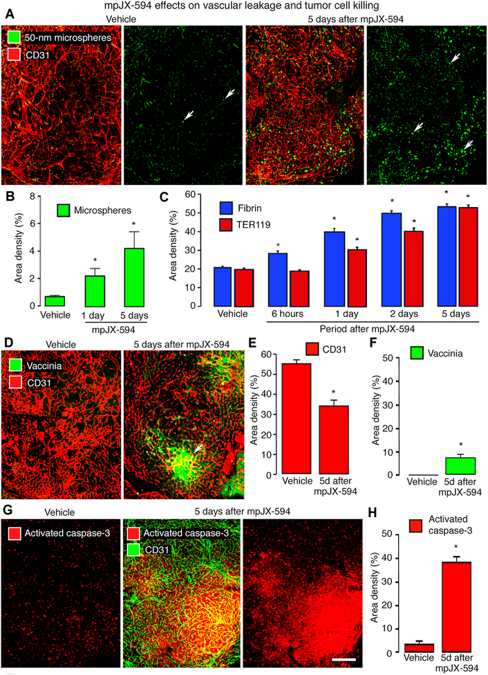
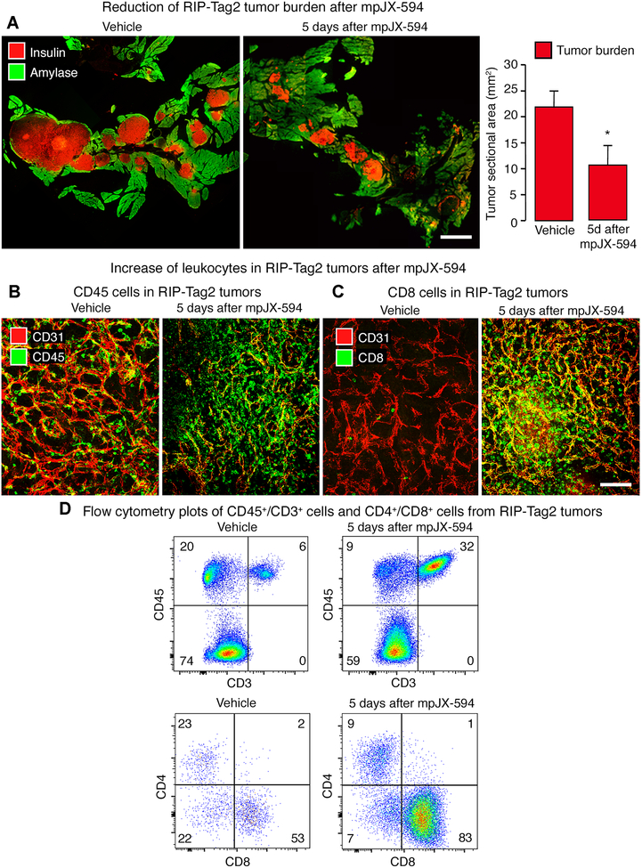
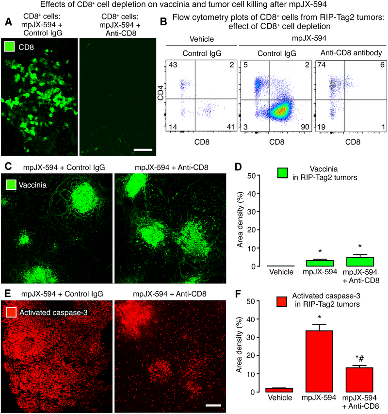
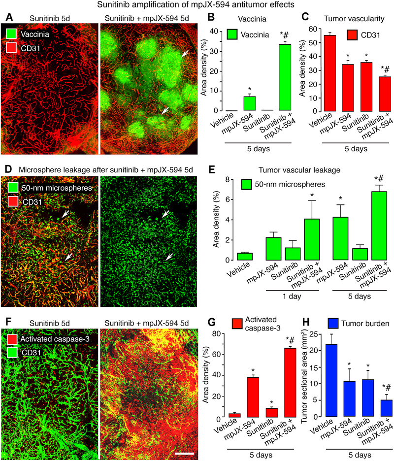
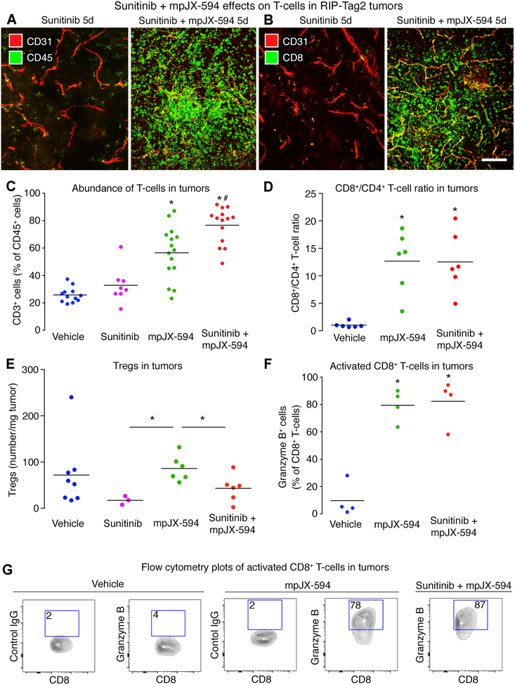
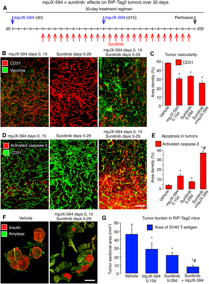
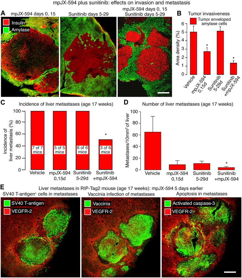
Oncolytic viruses pose many questions in their use in cancer therapy. In this study, we assessed the potential of mpJX-594 (mouse-prototype JX-594), a replication-competent vaccinia virus administered by intravenous injection, to target the tumor vasculature, produce immune activation and tumor cell killing more widespread than the infection, and suppress invasion and metastasis. These actions were examined in RIP-Tag2 transgenic mice with pancreatic neuroendocrine tumors that developed spontaneously and progressed as in humans. mpJX-594 initially infected tumor vascular endothelial cells, leading to vascular pruning and prolonged leakage in tumors but not in normal organs; parallel effects were observed in U87 gliomas. Viral infection spread to tumor cells, where tumor cell killing was much more widespread than the infection. Widespread tumor cell killing at 5 days was prevented by depletion of CD8+ T lymphocytes and did not require GM-CSF, as mpJX-594 variants that expressed human, mouse, or no GM-CSF produced equivalent amounts of killing. The antivascular, antitumor, and antimetastatic effects of mpJX-594 were amplified by concurrent or sequential administration of sunitinib, a multitargeted receptor tyrosine kinase inhibitor. These effects were not mimicked by selective inhibition of VEGFR2 despite equivalent vascular pruning, but were accompanied by suppression of regulatory T cells and greater influx of activated CD8+ T cells. Together, our results showed that mpJX-594 targets tumor blood vessels, spreads secondarily to tumor cells, and produces widespread CD8+ T-cell-dependent tumor cell killing in primary tumors and metastases, and that these effects can be amplified by coadministration of sunitinib.Significance: These findings reveal multiple unrecognized features of the antitumor properties of oncolytic vaccinia viruses, all of which can be amplified by the multitargeted kinase inhibitor sunitinib. Cancer Res; 78(4); 922-37. ©2017 AACR.


Conflict of interest statement

Disclosure of Potential Conflicts of Interest: Caroline J. Breitbach, John Bell, and David H. Kirn are holders of SillaJen company stock or stock options.

Figures

Figure 1.
Figure 1.. mpJX-594 effects on vascular leakage and tumor cell killing.
A: Extravasated 50-nm microspheres (green), shown with blood vessels (CD31, red) and alone in RIP-Tag2 tumors, are sparse at 5 days after vehicle but are abundant after mpJX-594. B: Microsphere extravasation was greater at 5 days than at 1 day after mpJX-594. ANOVA. *P < 0.05 compared to vehicle (n = 4 mice/group). C: Time-dependent increase in staining for extravasated fibrin (blue) and erythrocytes (TER119, red) in tumors from 6 hours to 5 days after iv injection of mpJX-594. ANOVA. *P < 0.05 for difference compared to vehicle for 5 days (n = 5 mice/group). D: Confocal micrographs of blood vessels (CD31, red) and vaccinia staining (green, arrow) in RIP-Tag2 tumors at 5 days after one dose of vehicle or mpJX-594. E, F: Differences in tumor vascularity (CD31 staining) and vaccinia staining at 5 days after vehicle or mpJX-594. *P < 0.05 compared to vehicle (n = 5 mice/group). G: Confocal micrographs of apoptotic cells (activated caspase-3, red) stained alone and with blood vessels (CD31, green) in RIP-Tag2 tumors at 5 days after vehicle or mpJX-594. H: Large increase in activated caspase-3 staining at 5 days after mpJX-594. ANOVA. *P < 0.05 compared to vehicle (n = 5 mice/group). Scale bar in G applies to all images: 200 μm.
Figure 2.
Figure 2.. mpJX-594 effects on tumor burden and leukocyte influx.
A: Tumor size indicated by staining of tumor cells for insulin (red) and pancreatic acinar cells for amylase (green) at 5 days after one dose of vehicle (left) or mpJX-594 (right). Graph shows corresponding measurements of tumor burden estimated as combined area of all tumors in sections of pancreas (see Methods). Student’s t-test. *P < 0.05 compared to vehicle (n = 5 mice/group). B: Confocal micrographs of leukocytes (CD45, green) and blood vessels (CD31, red) in RIP-Tag2 tumors at 5 days after vehicle or mpJX-594. C: Confocal micrographs show greater abundance of CD8+ T-cells (green) in tumors after mpJX-594. Blood vessels (CD31, red). D: Flow cytometry plots of CD45+/CD3+ cells (from all living cells in tumor) and CD4+/CD8+ cells (from CD45+/CD3+ fraction) isolated from tumors show increases in CD3+ cells and CD8+ cells (percentages in corners) at 5 days after mpJX-594. Scale bar 3 mm in A and 100 μm in C.
Figure 3.
Figure 3.. Effects of CD8+ T-cell depletion on vaccinia and apoptosis after mpJX-594.
A: Confocal micrographs of CD8+ T-cells in RIP-Tag2 tumors at 5 days after mpJX-594 in mice with intact CD8+ T-cells (left) or after CD8+ T-cell depletion (right) by anti-CD8 antibody. B: Flow cytometry plots of T-cells (pre-gated on CD45+/CD3+) compare the small CD8+ T-cell population after vehicle (left panel) to the large population at 5 days after mpJX-594 (center panel). CD8+ T-cells were not detected after mpJX-594 accompanied by CD8+ T-cell depletion (right panel). C: Confocal micrographs of vaccinia staining in RIP-Tag2 tumor sections after mpJX-594 given with control IgG or anti-CD8 antibody as in B. D: Amounts of vaccinia staining after mpJX-594 were similar with or without CD8+ T-cell depletion. Vaccinia was absent in vehicle-treated controls. E: Confocal micrographs of activated-caspase-3 staining in tumor sections adjacent to those stained for vaccinia. Apoptotic cells in RIP-Tag2 tumors after mpJX-594 with anti-CD8+ antibody are less abundant than with control IgG and are concentrated in regions of vaccinia staining. F: Greater activated caspase-3 staining after mpJX-594 but significantly less when virus was accompanied by CD8+ T-cell depletion. ANOVA. P < 0.05 compared to * vehicle or to # mpJX-594 plus control IgG (n = 3–6 mice/group). Scale bars 50 μm in A and 200 μm in E.
Figure 4.
Figure 4.. Sunitinib amplification of mpJX-594 effects on tumor vascularity, leakage, and apoptosis.
A: Confocal micrographs of blood vessels (CD31, red) and vaccinia (green, arrows) in RIP-Tag2 tumors at 5 days after daily sunitinib or one dose of mpJX-594 plus daily sunitinib. B, C: Treatment-related differences in vaccinia (B) and blood vessels (C) in tumors. ANOVA. P < 0.05 compared to * vehicle or to # mpJX-594 alone (n = 5 mice/group). D: Widespread extravasation of 50-nm microspheres (green, arrows) in tumors at 5 days after mpJX-594 plus sunitinib. Intravascular microspheres removed by vascular perfusion 10 minutes after iv injection of tracer. Blood vessels (CD31, red). E: Extravasated microspheres were more abundant at 5 days after mpJX-594 than vehicle and were more numerous after mpJX-594 plus sunitinib than mpJX-594 alone. P < 0.05 compared to * vehicle or to # mpJX-594 alone (n = 4 mice/group). F: Confocal micrographs show more apoptotic cells (activated caspase-3, red) in RIP-Tag2 tumor at 5 days after mpJX-594 plus sunitinib than sunitinib alone. Blood vessels (CD31, green). G, H: Treatment-related differences in activated caspase-3 (G) and tumor burden in pancreas (H). P < 0.05 compared to * vehicle or to # mpJX-594 alone (n = 5 mice/group). Scale bar in F applies to all images: 200 μm.
Figure 5.
Figure 5.. CD8+ T-cell influx after mpJX-594 plus sunitinib.
A, B: Confocal micrographs of leukocytes stained for CD45 (A, green) or CD8 (B, green) and blood vessels (CD31, red) in RIP-Tag2 tumors at 5 days after sunitinib alone or with mpJX-594. C: Treatment-related differences in proportions of CD3+ cells in CD45+ leukocyte population assessed by flow cytometry. Each dot represents value for all tumors from one RIP-Tag2 mouse. ANOVA. P < 0.05 compared to * vehicle or to # mpJX-594 alone (n = 8–15 mice/group). D: Ratio of CD8+ T-cells to CD4+ T-cells in tumors from mice at 5 days after vehicle, mpJX-594, or mpJX-594 plus sunitinib. ANOVA. *P < 0.05 compared to vehicle (n = 6 mice/group). E: Treatment-related differences in regulatory T cells (Tregs, CD45+/CD3+/CD4+/Foxp3+) per mg tumor tissue at 5 days. No significant difference among groups by ANOVA, but *P < 0.05 for differences between sunitinib or sunitinib plus mpJX-594 versus mpJX-594 alone by Student’s t-tests (n = 3–8 mice/group). F: Treatment-related differences in CD8+ T-cell activation shown by flow cytometry for granzyme B+ staining. ANOVA. *P < 0.05 compared to vehicle (n = 4 mice/group). G: Flow cytometry plots show gating and upregulation of granzyme B in CD8+ T-cells at 5 days after mpJX-594 alone or with sunitinib (% granzyme B+ cells in boxes, pre-gated on CD45+/CD3+/CD8+). Control antibody is mouse IgG of same isotype. Scale bar 100 μm in B.
Figure 6.
Figure 6.. mpJX-594 plus sunitinib: 30-day treatment of RIP-Tag2 mice.
A: Treatment regimens for mpJX-594 (2 doses, days 0, 15) and sunitinib (25 doses, days 5–29) individually or together. Controls received vehicle for 30 days. B: Confocal micrographs show treatment-related differences in vaccinia (green) and blood vessels (CD31, red) in tumors. C: Treatment-related differences in tumor vascularity. D: Confocal micrographs compare abundance of apoptotic cells (activated caspase-3, red) and blood vessels (CD31, green) after three treatments. E: Treatment-related differences activated caspase-3 in tumors. F: Tumor cells (insulin, red) and pancreatic acinar cells (amylase, green) after treatment with vehicle (left) or mpJX-594 plus sunitinib over 30 days (right). G: Treatment-related differences in tumor burden assessed as total sectional area of SV40 T-antigen stained tumors in the pancreas. ANOVA for comparisons in C, E, G. P < 0.05 compared to * vehicle or to # mpJX-594 alone or sunitinib alone (n = 5 mice/group). Scale bar in F applies to all images: 200 μm in B, D, and 3 mm in F.
Figure 7.
Figure 7.. mpJX-594 plus sunitinib: Effects on invasion and metastasis.
A: Confocal micrographs show treatment-related differences in invasion of tumor cells (insulin, red) into acinar pancreas (amylase, green) of RIP-Tag2 mice (treatment regimens in Fig. 7A). Tumor treated with sunitinib for 25 days is the most invasive, reflected by highly irregular border contour. B: Treatment-related differences in amount of invasion indicated by tumor cells surrounding acinar cells. ANOVA. *P < 0.05 compared to vehicle (n = 5 mice/group). C: Treatment-related differences in incidence of liver micrometastases. Number of mice with metastases shown on bars. Fisher’s exact test. *P = 0.0247. D: Treatment-related differences in abundance of liver micrometastases expressed per 10 mm2 of section area. ANOVA. *P < 0.05 compared to vehicle (n = 5 mice/group). E: Confocal micrographs of liver of 17-week-old mouse 5 days after iv injection of mpJX-594. Sequential sections stained for blood vessels (VEGFR-2, red) and tumor cells (SV40 T-antigen, green, left), vaccinia (green, center), or apoptotic cells (activated caspase-3, green, right). Scale bar in E applies to all images: 300 μm for A, 200 μm for E.

References

    1. Zeh HJ, Downs-Canner S, McCart JA, Guo ZS, Rao UN, Ramalingam L, et al. First-in-man study of western reserve strain oncolytic vaccinia virus: safety, systemic spread, and antitumor activity. Mol Ther 2015;23:202–14. - PMC - PubMed
    1. Puzanov I, Milhem MM, Minor D, Hamid O, Li A, Chen L, et al. Talimogene Laherparepvec in Combination With Ipilimumab in Previously Untreated, Unresectable Stage IIIB-IV Melanoma. J Clin Oncol 2016;34:2619–26. - PMC - PubMed
    1. Breitbach CJ, Lichty BD, Bell JC. Oncolytic Viruses: Therapeutics With an Identity Crisis. EBioMedicine 2016;9:31–6. - PMC - PubMed
    1. Lawler SE, Speranza MC, Cho CF, Chiocca EA. Oncolytic Viruses in Cancer Treatment: A Review. JAMA Oncol 2017;3:841–49. - PubMed
    1. Ribas A, Dummer R, Puzanov I, VanderWalde A, Andtbacka RHI, Michielin O, et al. Oncolytic Virotherapy Promotes Intratumoral T Cell Infiltration and Improves Anti-PD-1 Immunotherapy. Cell 2017;170:1109–19 e10. - PMC - PubMed

Publication types