. 2017 Dec 19;8(6):e01685-17.
doi: 10.1128/mBio.01685-17.

A Specialized Peptidoglycan Synthase Promotes Salmonella Cell Division inside Host Cells

Affiliations

A Specialized Peptidoglycan Synthase Promotes Salmonella Cell Division inside Host Cells

Sónia Castanheira et al. mBio. .

Abstract

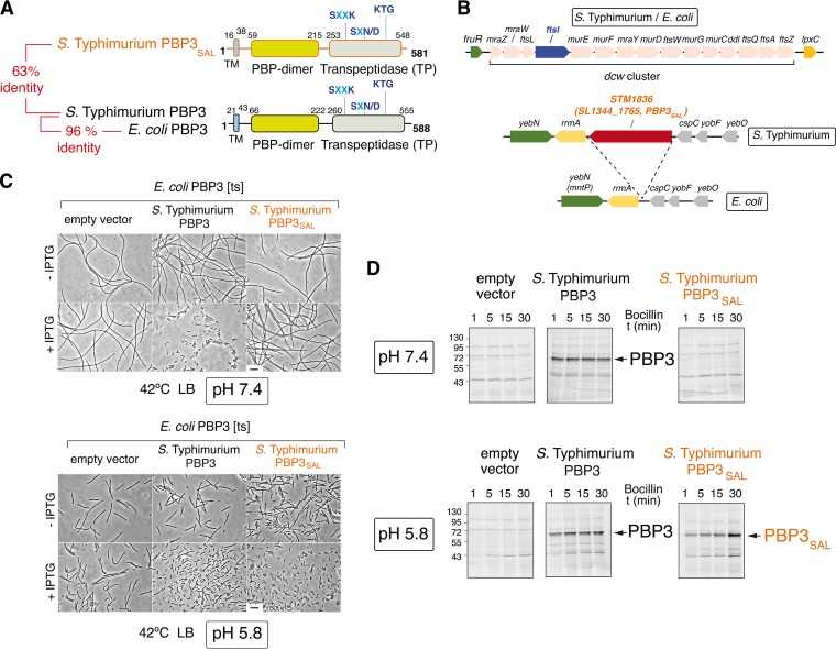
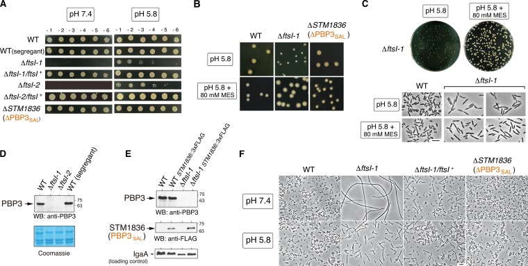
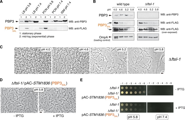
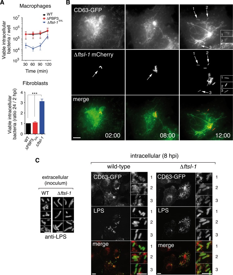
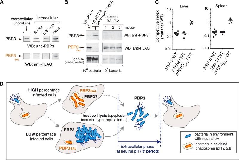
Bacterial cell division has been studied extensively under laboratory conditions. Despite being a key event in the bacterial cell cycle, cell division has not been explored in vivo in bacterial pathogens interacting with their hosts. We discovered in Salmonella enterica serovar Typhimurium a gene absent in nonpathogenic bacteria and encoding a peptidoglycan synthase with 63% identity to penicillin-binding protein 3 (PBP3). PBP3 is an essential cell division-specific peptidoglycan synthase that builds the septum required to separate daughter cells. Since S Typhimurium carries genes that encode a PBP3 paralog-which we named PBP3SAL-and PBP3, we hypothesized that there are different cell division events in host and nonhost environments. To test this, we generated S Typhimurium isogenic mutants lacking PBP3SAL or the hitherto considered essential PBP3. While PBP3 alone promotes cell division under all conditions tested, the mutant producing only PBP3SAL proliferates under acidic conditions (pH ≤ 5.8) but does not divide at neutral pH. PBP3SAL production is tightly regulated with increased levels as bacteria grow in media acidified up to pH 4.0 and in intracellular bacteria infecting eukaryotic cells. PBP3SAL activity is also strictly dependent on acidic pH, as shown by beta-lactam antibiotic binding assays. Live-cell imaging microscopy revealed that PBP3SAL alone is sufficient for S Typhimurium to divide within phagosomes of the eukaryotic cell. Additionally, we detected much larger amounts of PBP3SAL than those of PBP3 in vivo in bacteria colonizing mouse target organs. Therefore, PBP3SAL evolved in S Typhimurium as a specialized peptidoglycan synthase promoting cell division in the acidic intraphagosomal environment.IMPORTANCE During bacterial cell division, daughter cells separate by a transversal structure known as the division septum. The septum is a continuum of the cell wall and therefore is composed of membrane(s) and a peptidoglycan layer. To date, actively growing bacteria were reported to have only a "cell division-specific" peptidoglycan synthase required for the last steps of septum formation and consequently, essential for bacterial life. Here, we discovered that Salmonella enterica has two peptidoglycan synthases capable of synthesizing the division septum. One of these enzymes, PBP3SAL, is present only in bacterial pathogens and evolved in Salmonella to function exclusively in acidic environments. PBP3SAL is used preferentially by Salmonella to promote cell division in vivo in mouse target organs and inside acidified phagosomes. Our data challenge the concept of only one essential cell division-specific peptidoglycan synthase and demonstrate that pathogens can divide in defined host locations using alternative mechanisms.

Keywords: Salmonella; cell division; intracellular pathogens; penicillin-binding proteins; peptidoglycan.


Figures

FIG 1
FIG 1
The S. Typhimurium PBP3 paralog (PBP3SAL) is a division-specific peptidoglycan (PG) synthase active at acidic pH. (A) Functional domains and catalytic motifs of PBP3SAL, a protein absent in nonpathogenic bacteria, compared to those of PBP3 from S. Typhimurium and E. coli. TM, transmembrane region. Numbers indicate relative positions in the protein in which the TM and the functional domains are predicted. (B) S. Typhimurium genome regions harboring the ftsI and SL1344_1765 (STM1836) genes that encode PBP3 and PBP3SAL, respectively. The equivalent regions from the E. coli genome are shown, with complete synteny for ftsI and its flanking genes. Note that ftsI forms part of the dcw (division and cell wall gene cluster) operon and the S. Typhimurium SL1344_1765 (STM1836) gene inserted between rrmA and cspC, encoding a predicted 23S rRNA-methyltransferase and a cold shock protein-like protein, respectively. (C) At acidic pH (5.8), PBP3SAL restores division at 42°C in an E. coli ftsI(Ts) mutant that produces a heat-labile PBP3 variant (18). Noninducing (−IPTG) and inducing (+IPTG) expression conditions are shown. Bar, 5 µm. (D) PBP3SAL binds the beta‑lactam analog Bocillin-FL at acidic pH (5.8) but not neutral pH (7.4). Fluorescent assay showing Bocillin binding to membranes from E. coli strain RP41 [ftsI(Ts)] harboring an empty vector or vectors expressing S. Typhimurium PBP3 or PBP3SAL. Bocillin binding was analyzed at the pH in which bacteria were grown, either pH 5.8 or pH 7.4, as indicated. Numbers on the left of the gels refer to the molecular mass (in kDa) of the markers used. t, time.
FIG 2
FIG 2
Lack of PBP3 renders S. Typhimurium unable to grow at neutral pH (7.4) but retaining optimal growth at acidic pH. (A) Colony-forming capacity at pH 7.4 or 5.8 of the two clones lacking PBP3 (ΔftsI-1 and ΔftsI-2) obtained after segregation of the ftsI+ftsI merodiploid (see Fig. S3 in the supplemental material). Control strains include an ftsI+ (wild-type [WT]) segregant, the ΔftsI-1/ftsI+ and ΔftsI-2/ftsI+ complemented strains, and a ΔPBP3SAL mutant. Numbers on the top of the upper panels refer to serial dilutions of the bacterial culture (10-fold each successive dilution). (B) Growth of S. Typhimurium lacking PBP3 is further stimulated in acidic pH (5.8) LB medium buffered with 80 mM HEPES. Note that in buffered medium the mutant lacking PBP3 forms colonies even larger than those of the wild type and the mutant lacking PBP3SAL. (C) Despite differences in colony size in nonbuffered and buffered LB solid media (pH 5.8), the ΔftsI-1 mutant displays similar morphology and capacity for cell division at mid-exponential phase (OD600 of ~0.2 to 0.3) in nonbuffered and buffered LB liquid media (pH 5.8). (D) Western blot (WB) assays confirm the absence of PBP3 in membrane fractions obtained from the ΔftsI-1 and ΔftsI-2 mutants. (E) Unaltered relative PBP3SAL levels in the absence of PBP3, discarding coregulation between both proteins. The inner membrane protein IgaA (41) was used as a loading control. (F) The lack of cultivability of the ΔftsI-1 mutant at neutral pH (7.4) is due to a defect in cell division.
FIG 3
FIG 3
The amount of PBP3SAL produced by S. Typhimurium controls cell division in acidic environments. (A) Relative levels of PBP3 and PBP3SAL detected by Western blotting (WB) in total protein fraction of S. Typhimurium strain MD2559 [STM1836(PBP3SAL)::3×FLAG] grown at the indicated pH and growth phases in different laboratory media (LB, PCN [47], and ISM [48]). Note that the production of PBP3SAL is stimulated in acidified media. The results shown are representative of three independent experiments. (B) The relative levels of PBP3SAL increase as the pH of the media drops in the range of 5.8 to 4.0. Total protein fractions prepared at exponential phase (OD600 of ~0.2 to 0.3) in LB medium at the indicated pH values are shown. The outer membrane protein OmpA was used as a loading control. The results shown are representative of three experiments. (C) Cell division rate in the ΔftsI-1 mutant increases at lower pH values in concordance with higher levels of PBP3SAL. Note the gradual reduction in cell size as the pH of the medium decreases. (D) Increased cell division rate of the ΔftsI-1 mutant at pH 5.8 resulting from ectopic expression of PBP3SAL from plasmid. Note the reduction in cell size in inducing conditions (+ IPTG). (E) An increase in PBP3SAL relative levels does not restore cell division in neutral pH, supporting the specialization of PBP3SAL to function only in acidic environments. Numbers on the top of the upper panels refer to serial dilutions of the bacterial culture (10-fold each successive dilution).
FIG 4
FIG 4
PBP3SAL promotes division of intracellular S. Typhimurium. (A) Intracellular proliferation rates of isogenic mutants lacking PBP3 or PBP3SAL. Note the higher proliferation rate observed for the ΔftsI-1 mutant defective in PBP3. Data are the mean ± standard error (error bars) from three independent experiments. Values that are significantly different (P < 0.001) are indicated (***). (B) Time-lapse microscopy of live cells shows proliferation of intracellular ΔftsI-1 bacteria in fibroblast phagosomes. Transfected NRK-49F fibroblasts stably expressing the phagosomal membrane glycoprotein CD63 (34) were used. Images correspond to the same infected fibroblast, acquired at the indicated times (in hours) postinfection. The arrows and numbers (1 to 3) indicate different intracellular bacteria within phagosomal compartments. Bar, 10 µm. (C) Restoration of normal cell size in the ΔftsI-1 mutant when bacteria colonize the acidic intraphagosomal environment in fibroblasts. Bars, 5 µm (extracellular), 10 µm (intracellular wild-type and ΔftsI-1 strains), 5 µm (insets for wild-type and ΔftsI-1 strains). The numbers 1, 2, and 3 in the insets for intracellular wild-type and ΔftsI-1 strains show different fields of the same image.
FIG 5
FIG 5
S. Typhimurium increases PBP3SAL production inside eukaryotic cells and in vivo in mouse organs. (A) Relative levels of PBP3 and PBP3SAL detected in intracellular bacteria (PBP3SAL::3×FLAG-tagged strain) after infection of BJ-5ta human fibroblasts (24 hpi), NRK-49F rat fibroblasts (24 hpi), and HeLa human epithelial cells (16 hpi). Extracellular bacteria used to infect the cells (inoculum grown in LB at pH 7.4) were used as a control. The results shown are representative of three independent experiments. (B) PBP3SAL production increases at higher levels than PBP3 in vivo. The relative levels of both proteins in spleen extracts from three BALB/c mice challenged intraperitoneally with the PBP3SAL::3×FLAG-tagged strain are shown. (C) Competitive index between isogenic mutants lacking either PBP3 or PBP3SAL and wild-type bacteria following intraperitoneal challenge of BALB/c mice. Liver and spleen extracts were obtained at 48 h postchallenge. (D) Model depicting the contribution of PBP3 and PBP3SAL to S. Typhimurium cell division in different host environments: intracellular (neutral pH cytosol or acidic phagosomes) and extracellular sites during transit between infected cells. Note that the absence of PBP3 can impair normal progression of the infection, since bacteria necessarily pass an extracellular stage in a neutral pH environment (referred to here as the “t” period). A long “t” period could result for ΔftsI bacteria in long filamentous cells (Fig. 2F), unable to infect new nearby host cells. In contrast, the versatile PBP3 could promote division in the phagosomal environment in the absence of PBP3SAL.

Comment in

References

    1. Adams DW, Errington J. 2009. Bacterial cell division: assembly, maintenance and disassembly of the Z ring. Nat Rev Microbiol 7:642–653. doi: 10.1038/nrmicro2198. - DOI - PubMed
    1. Lutkenhaus J, Pichoff S, Du S. 2012. Bacterial cytokinesis: from Z ring to divisome. Cytoskeleton 69:778–790. doi: 10.1002/cm.21054. - DOI - PMC - PubMed
    1. Söderström B, Daley DO. 2017. The bacterial divisome: more than a ring? Curr Genet 63:161–164. doi: 10.1007/s00294-016-0630-2. - DOI - PubMed
    1. Tsang MJ, Bernhardt TG. 2015. Guiding divisome assembly and controlling its activity. Curr Opin Microbiol 24:60–65. doi: 10.1016/j.mib.2015.01.002. - DOI - PMC - PubMed
    1. Harry EJ. 2001. Bacterial cell division: regulating Z-ring formation. Mol Microbiol 40:795–803. doi: 10.1046/j.1365-2958.2001.02370.x. - DOI - PubMed

Publication types

LinkOut - more resources