. 2018 Feb 9;293(6):1875-1886.
doi: 10.1074/jbc.RA117.000754. Epub 2017 Dec 19.

Stepwise processing analyses of the single-turnover PCSK9 protease reveal its substrate sequence specificity and link clinical genotype to lipid phenotype

Affiliations

Stepwise processing analyses of the single-turnover PCSK9 protease reveal its substrate sequence specificity and link clinical genotype to lipid phenotype

John S Chorba et al. J Biol Chem. .

Erratum in

Abstract

Proprotein convertase subtilisin/kexin type 9 (PCSK9) down-regulates the low-density lipoprotein (LDL) receptor, elevating LDL cholesterol and accelerating atherosclerotic heart disease, making it a promising cardiovascular drug target. To achieve its maximal effect on the LDL receptor, PCSK9 requires autoproteolysis. After cleavage, PCSK9 retains its prodomain in the active site as a self-inhibitor. Unlike other proprotein convertases, however, this retention is permanent, inhibiting any further protease activity for the remainder of its life cycle. Such inhibition has proven a major challenge toward a complete biochemical characterization of PCSK9's proteolytic function, which could inform therapeutic approaches against its hypercholesterolemic effects. To address this challenge, we employed a cell-based, high-throughput method using a luciferase readout to evaluate the single-turnover PCSK9 proteolytic event. We combined this method with saturation mutagenesis libraries to interrogate the sequence specificities of PCSK9 cleavage and proteolysis-independent secretion. Our results highlight several key differences in sequence identity between these two steps, complement known structural data, and suggest that PCSK9 self-proteolysis is the rate-limiting step of secretion. Additionally, we found that for missense SNPs within PCSK9, alterations in both proteolysis and secretion are common. Last, we show that some SNPs allosterically modulate PCSK9's substrate sequence specificity. Our findings indicate that PCSK9 proteolysis acts as a commonly perturbed but critical switch in controlling lipid homeostasis and provide a new hope for the development of small-molecule PCSK9 inhibitors.

Keywords: active site; atherosclerosis; high-throughput screening (HTS); low-density lipoprotein (LDL); proprotein convertase subtilisin/kexin type 9 (PCSK9); protein secretion; serine protease; single-nucleotide polymorphism (SNP); substrate specificity.


Conflict of interest statement

The authors declare that they have no conflicts of interest with the contents of this article

Figures

Figure 1.
Figure 1.
PCSK9 cleavage assay and mutagenesis library. A, schematic of the in trans PCSK9 proteolysis assay. Co-expression of the substrate (Prodomain-NLuc) with a non-secretable prodomain-deficient PCSK9 protease (PCSK9ΔPro-C679X) allows for secretion of the luciferase, and a positive readout in the conditioned medium, only when cleavage is permitted. In this situation, the system perturbation is a library of cleavage site mutants as substrates. See “Results” for further details. B, relative proteolysis of WT versus inactive (S386A) PCSK9 protease. Error bars, S.D. Results of an unpaired t test with Welch's correction and the Z′ factor (62) are shown. C, schematic for design of a saturation mutagenesis library of proteolysis substrates. The design of the paired oligonucleotides to replace the BsaI cleavage site shown reflects the WT sequence, although each codon was sequentially exchanged for the NNK equivalent to generate the library. See “Results” for details.
Figure 2.
Figure 2.
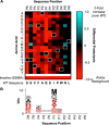
PCSK9 cleavage sequence specificity. A, heat map showing the relative cleavage for each single amino acid mutant in the P6–P6′ cleavage sequence as compared with the WT sequence. White rectangles, WT amino acid. A value of −1 (red) indicates essentially no proteolysis, and a value of +1 (cyan) indicates a 2-fold increase from the WT value. Mutants with statistically significant differences from WT, as determined by a Holm–Sidak corrected unpaired t test with α < 0.05, are shown in color. Mutants with effects not statistically different are shown in black. B, WebLogo (27) illustrating the preferred cleavage sequence for PCSK9. The WT amino acid is shown outlined in black, with other residues colored to reflect the values in the heat map.
Figure 3.
Figure 3.
Crystal structure of the PCSK9 active site and bound prodomain tail. Shown is the structure of mature PCSK9 (Protein Data Bank code 2P4E) (12) with missing internal loops optimized by ModLoop (63). The active site with the bound prodomain C terminus (magenta, residues 147–152) is magnified, and the residues are identified. The right panel shows a cut-away view of the prodomain residues protruding directly into the binding groove. See “Results” for discussion.
Figure 4.
Figure 4.
PCSK9 secretion assay and mutagenesis library. A, schematic of the in trans PCSK9 secretion assay. Co-expression of the prodomain with a luciferase-tagged, prodomain-deficient PCSK9 (PCSK9ΔPro-NLuc) allows for readout of the luciferase in the conditioned medium only when secretion of the PCSK9 is permitted. In this situation, the system perturbation is a library of prodomain tail mutants. See “Results” for details. B, relative proteolysis of WT versus non-secreted (S462P) PCSK9 protease. Error bars, S.D. Results of an unpaired t test with Welch's correction and the Z′ factor (62) are shown. C, schematic for design of saturation mutagenesis library of secretory prodomain tail mutants. The design of the paired oligonucleotides to replace the BsaI cleavage site shown reflects the WT sequence, although each codon was sequentially exchanged for the NNK equivalent to generate the library. See “Results” for details.
Figure 5.
Figure 5.
PCSK9 secretion sequence specificity. A, heat map showing the relative secretion for each single amino acid mutant in the P6–P1 cleavage sequence as compared with the WT sequence. White rectangles, WT amino acid. A value of −1 (red) indicates essentially no secretion, and a value of +1 (cyan) indicates a 2-fold increase from the WT value. Mutants with statistically significant differences from WT, as determined by Holm–Sidak corrected unpaired t test with α < 0.05, are shown in color. Mutants with effects not statistically different are shown in black. B, WebLogo (27) illustrating the preferred secretion sequence for PCSK9. The WT amino acid is shown outlined in black, with other residues colored to reflect the values in the heat map. C, heat map showing the differential cleavage for the single-amino acid mutants in the proteolysis versus secretion assays. Mutants with statistically significant differences between proteolysis and secretion assays, as determined by Holm–Sidak corrected unpaired t test with α < 0.05, are shown in color. Mutants with effects not statistically different are shown in black. Values in green illustrate preferential proteolysis, whereas those in cyan illustrate preferential secretion.
Figure 6.
Figure 6.
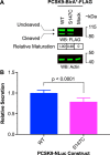
Cleavage is the rate-limiting step of PCSK9 processing. A, immunoblot of cell lysates transiently expressing PCSK9-BirA*-FLAG WT or S147C constructs. The relative maturation, as quantified on the immunoblot, is noted below the appropriate column. B, relative secretion of full-length PCSK9 WT versus S147C, as measured by luciferase activity. Error bars, S.D. Results of an unpaired t test with Welch's correction are shown.
Figure 7.
Figure 7.
Clinical SNPs frequently affect PCSK9 processing. Top, individual SNPs chosen for analysis graphically represented as yellow bars with black outlines on the protein primary structure, with PCSK9 domains noted above. Residues with two SNPs at a given position are denoted by two half-height bars stacked vertically. Middle, SNPs with statistically significant differences in proteolysis compared with WT, as determined by Holm–Sidak corrected unpaired t test with α < 0.05. A value of −1 (red) indicates essentially no processing activity, and a value of +1 (cyan) indicates a 2-fold improvement over WT. Bottom, SNPs with statistically significant differences in secretion compared with WT, as determined by a Holm–Sidak corrected unpaired t test with α < 0.05. The color scheme is the same as for the proteolysis assay.
Figure 8.
Figure 8.
Allosteric effects of PCSK9 SNPs. A, crystal structure of mature PCSK9 (Protein Data Bank code 2P4E) (12) with missing internal loops optimized by ModLoop (63), with selected SNPs with improved proteolytic ability labeled in green. B, effect of individual SNPs on proteolytic activity of selected cleavage sequence mutants. To compare the effect of the SNPs on sequence specificity, values are normalized to the activity of that SNP on the WT cleavage sequence. Error bars, S.D. Results of unpaired t tests with Welch's correction are shown. C, effect of optimized cleavage sequence (SSVFAQS) replacing the native 212–220 (GTRFHRQA) sequence in full-length PCSK9-NanoLuc on relative secretion. Error bars, S.D. Results of an unpaired t test with Welch's correction are shown.

References

    1. Park S. W., Moon Y. A., and Horton J. D. (2004) Post-transcriptional regulation of low density lipoprotein receptor protein by proprotein convertase subtilisin/kexin type 9a in mouse liver. J. Biol. Chem. 279, 50630–50638 10.1074/jbc.M410077200 - DOI - PubMed
    1. Maxwell K. N., Fisher E. A., and Breslow J. L. (2005) Overexpression of PCSK9 accelerates the degradation of the LDLR in a post-endoplasmic reticulum compartment. Proc. Natl. Acad. Sci. U.S.A. 102, 2069–2074 10.1073/pnas.0409736102 - DOI - PMC - PubMed
    1. Zhang D.-W., Lagace T. A., Garuti R., Zhao Z., McDonald M., Horton J. D., Cohen J. C., and Hobbs H. H. (2007) Binding of proprotein convertase subtilisin/kexin type 9 to epidermal growth factor-like repeat A of low density lipoprotein receptor decreases receptor recycling and increases degradation. J. Biol. Chem. 282, 18602–18612 10.1074/jbc.M702027200 - DOI - PubMed
    1. Grefhorst A., McNutt M. C., Lagace T. A., and Horton J. D. (2008) Plasma PCSK9 preferentially reduces liver LDL receptors in mice. J. Lipid Res. 49, 1303–1311 10.1194/jlr.M800027-JLR200 - DOI - PMC - PubMed
    1. Seidah N. G. (2016) New developments in proprotein convertase subtilisin–kexin 9's biology and clinical implications. Curr. Opin. Lipidol. 27, 274–281 10.1097/MOL.0000000000000295 - DOI - PubMed

Publication types

Associated data