. 2017 Dec 19;8(1):2196.
doi: 10.1038/s41467-017-02109-8.

Unravelling the specificity and mechanism of sialic acid recognition by the gut symbiont Ruminococcus gnavus

Affiliations

Unravelling the specificity and mechanism of sialic acid recognition by the gut symbiont Ruminococcus gnavus

C David Owen et al. Nat Commun. .

Abstract

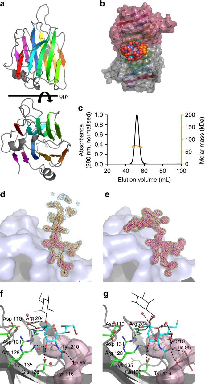
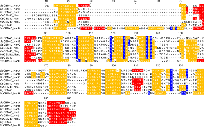
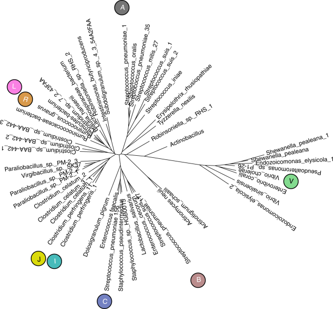
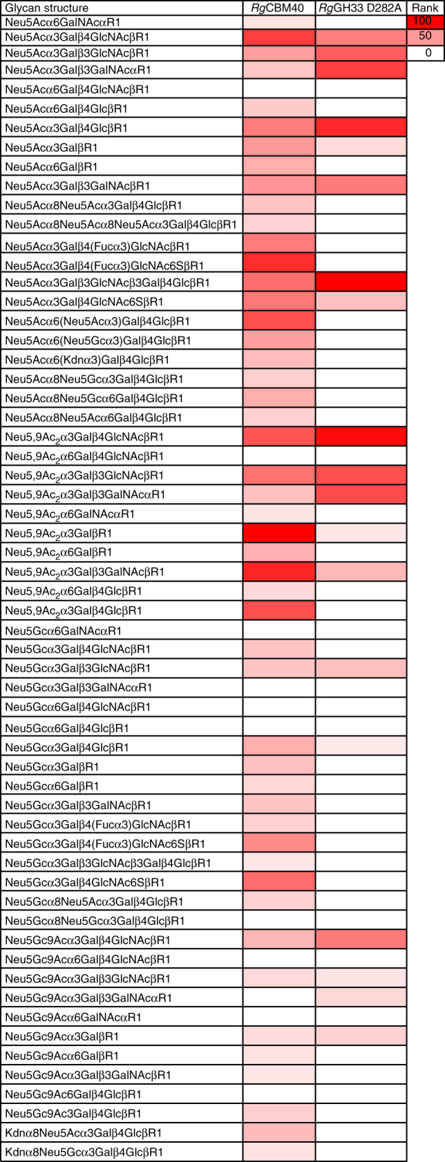
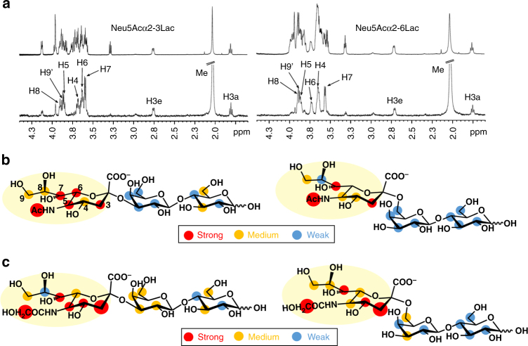
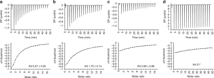
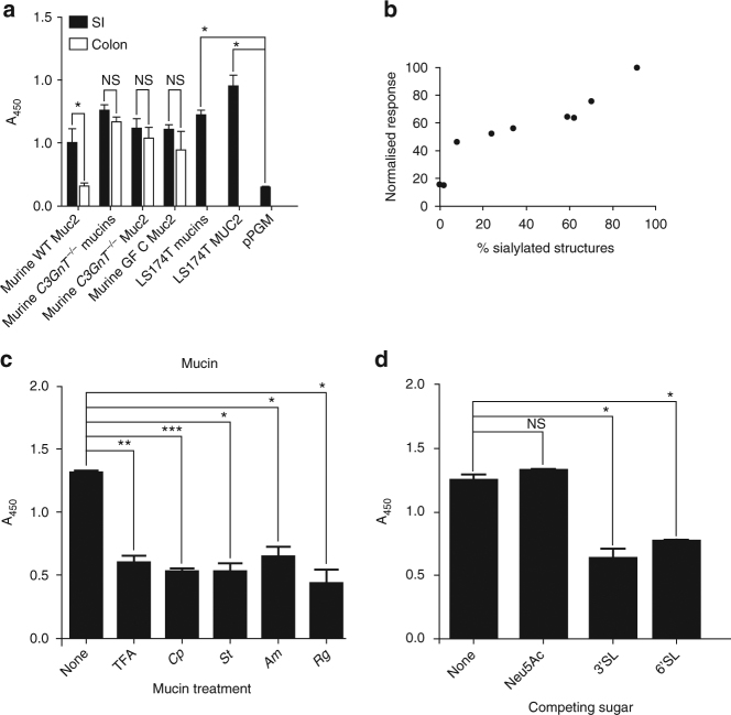
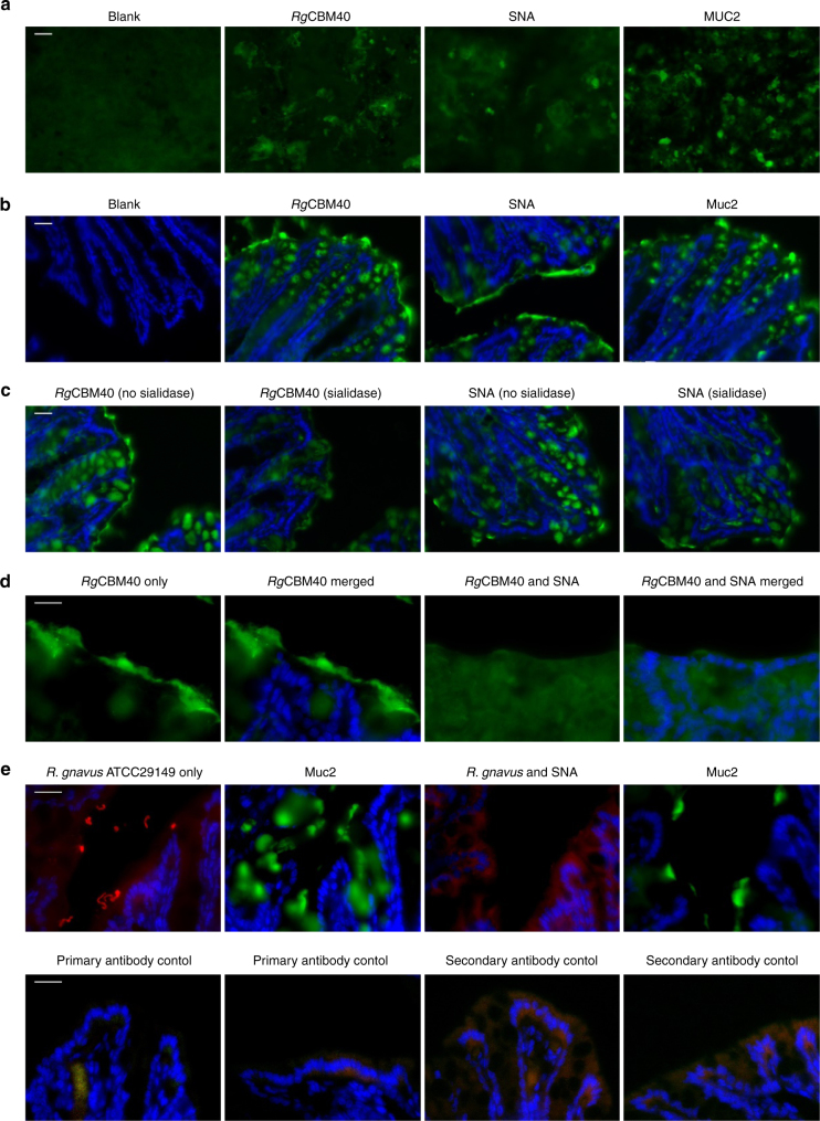
Ruminococcus gnavus is a human gut symbiont wherein the ability to degrade mucins is mediated by an intramolecular trans-sialidase (RgNanH). RgNanH comprises a GH33 catalytic domain and a sialic acid-binding carbohydrate-binding module (CBM40). Here we used glycan arrays, STD NMR, X-ray crystallography, mutagenesis and binding assays to determine the structure and function of RgNanH_CBM40 (RgCBM40). RgCBM40 displays the canonical CBM40 β-sandwich fold and broad specificity towards sialoglycans with millimolar binding affinity towards α2,3- or α2,6-sialyllactose. RgCBM40 binds to mucus produced by goblet cells and to purified mucins, providing direct evidence for a CBM40 as a novel bacterial mucus adhesin. Bioinformatics data show that RgCBM40 canonical type domains are widespread among Firmicutes. Furthermore, binding of R. gnavus ATCC 29149 to intestinal mucus is sialic acid mediated. Together, this study reveals novel features of CBMs which may contribute to the biogeography of symbiotic bacteria in the gut.


Conflict of interest statement

The authors declare no competing financial interests.

Figures

Fig. 1
Fig. 1
Crystal structure of RgCBM40 in complex with 3′SL and 6′SL. a RgCBM40 is shown in a cartoon representation with a rotation of 90° around the x axis. b The protein crystallised as a dimer with the ligand binding site at the dimer interface. The binding sites are shown occupied by 6′SL trisaccharides (Neu5Ac: cyan, galactose: blue, glucose: orange). c SEC-MALS performed with full length RgNanH (77 kDa). The SEC-MALS predicted molecular weight was 73 kDa, indicating that RgNanH is monomeric in solution. Bound (d) 3′SL and (e) 6′SL are shown with their corresponding Fo-Fc omit maps at 2σ (light cyan), 3σ (orange) and 5σ (magenta). The omit maps are carved at 1.6 Å around the bound ligand. For 3′SL, the map is carved around a dummy glucose residue to indicate the presence of partial electron density. A close-up view of RgCBM40 binding site is shown with (f) 3′SL and (g) 6′SL. The Neu5Ac residue is shown in cyan and the galactose residue as black lines, for clarity the glucose residue is not shown. Interacting RgCBM40 residues are shown in green with black dashed lines indicating hydrogen bonding interactions. A semi-transparent surface indicates the hydrophobic surface
Fig. 2
Fig. 2
CBM40 structural alignment. Structure-based alignment (α-helices and β-strands respectively in red and yellow) of CBM40 domains of RgCBM40 with C. perfringens CpCBM40_NanJ (PDB code 2V73) and CpCBM40_NanI (PDB code 5FRA), M. decora MdCBM40_NanL (PDB code 1SLI) and S. pneumoniae SpCBM40_NanA (PDB code 4C1W), SpCBM40_NanB (PDB code 2VW0) and SpCBM40_NanC (PDB code 4YZ5) and VcCBM40_NanH structure (PDB code 2W68). Amino acids identified as binding sites are highlighted in blue. RgCBM40 residues Ile95, Asp110, Tyr116, Glu126, Arg128, Arg204 and Tyr210 are at positions 104, 119, 125, 135, 137, 226 and 233 of the alignment. The alignment supplemented with other canonical and Vibrio-type CBM40 sequences, used to create the pHMM using HMMER3, is shown in Supplementary Fig. 4
Fig. 3
Fig. 3
Distance-based tree of canonical and Vibrio-type CBM40 sequences. Tree of 51 non-redundant sequences (80% identity level) calculated by neighbour-joining using evolutionary distances estimated by applying the PMB model of amino acid changes, including all sites and using a uniform rate of evolution. The representative sequences corresponding most closely (at least 97% identical) to the 7 bacterial structure-determined sequences are shown with symbols, coloured in accordance with Supplementary Fig. 1: “A”, SpCBM40_NanA; “B”, SpCBM40_NanB; “C”, SpCBM40_NanC; “I”, CpCBM40_NanI; “J”, CpCBM40_NanJ; “R”, RgCBM40; “V”, VcCBM40_NanH. Additionally, “L” denotes MdCBM40_NanL closest to the bacterial sequence of highest identity (70% identical to RgCBM40) as only bacterial sequences were searched
Fig. 4
Fig. 4
Sialoglycan microarray analysis of binding specificities of RgCBM40 and RgGH33 D282A. Binding of the recombinant proteins RgCBM40 and RgGH33 D282A at 20 and 200 µg ml−1, respectively are presented (n = 4, SD). Heat map was generated using the method as previously described,. Binding was ranked as (glycan average RFU/ maximum glycan average RFU)×100. Red and white represent the maximum and minimum, respectively. R1 represents propyl-azide as the spacer
Fig. 5
Fig. 5
STD NMR analysis of RgCBM40 binding to sialoglycans. a Reference (top) and difference (bottom) spectra of 3′SL and 6′SL. The strongest signals from the Neu5Ac’s protons are labelled in the difference spectra. b Binding epitope mapping from STD NMR of 3′SL and 6′SL. Legend indicates relative STD intensities normalised at H7: blue, 0–24%; yellow, 25–50%; red 51–100%; larger red dots indicate values over 100%. Sialic acid is the main recognition element. c Binding epitopes mapping from STD NMR of Neu5Gcα2-3Lac and Neu5Gcα2-6Lac. Legend as above. Sialic acid is the main recognition element. The strongest STD intensities from CH2 and the H3s, suggest a reorientation of the Neu5Gc ring in the binding pocket, in comparison to 3′SL and 6′SL
Fig. 6
Fig. 6
ITC isotherms of RgCBM40 to sialoglycans. a RgCBM40 binding to 3′SL, b RgCBM40 binding to 6′SL, c RgCBM40 binding to 3′SLGc, d RgCBM40 binding to Neu5Ac. The Kd is indicated in mM. *This value is an estimate as the Kd is too high to determine with the concentration of sugar used
Fig. 7
Fig. 7
ELISA of RgCBM40 binding to purified mucins. a RgCBM40 binding to a range of purified mucins; mucin 2 (MUC2) and mixed mucins (mucins) from human cell line LS174T, purified pig gastric mucin (pPGM), and murine mucins from germ free (GF), wild type (WT), and C3GnT −/− mice. b Correlation of RgCBM40 binding with % sialylated structure for each mucin tested. The % sialylated structures was determined by MS. c RgCBM40 binding to LS174T MUC2 which has been treated chemically (TFA) or enzymatically with a sialidase from Clostridium perfringens (Cp), Salmonella typhimurium (St), Akkermansia muciniphila (Am) or Ruminococcus gnavus (Rg) d RgCBM40 binding to LS174T MUC2 in competition with sugars. RgCBM40 has been preincubated with the indicated sugars. In all cases, RgCBM40 was incubated with immobilised mucins and binding detected using an anti-sialidase primary antibody and an anti-rabbit secondary antibody conjugated to horseradish peroxidase. The enzyme was incubated with TMB and the absorbance at 450 nm (A450) measured. The error bars show the standard error of the mean (SEM) of three replicates. P values are indicated; NS-not significant, *p < 0.05, **p < 0.005, ***p < 0.0005
Fig. 8
Fig. 8
RgCBM40 binding to mucus-producing cells and intestinal tissue sections. a Immunostaining pattern for RgCBM40 on LS174T cells correlated with mucin (MUC2) and lectin (SNA) staining, all shown in green. No staining was observed in RgCBM40-free sample (Blank). b Immunostaining pattern for RgCBM40 on cryosections of mouse colon correlated with mucin (Muc2) and lectin (SNA) staining, all shown in green. No staining was observed in RgCBM40-free sample (Blank). Cell nuclei were counterstained with DAPI, shown in blue. c Sialidase pre-treatment of mouse colonic cryosections markedly reduced the binding of RgCBM40 and SNA lectin. Cell nuclei were counterstained with DAPI, shown in blue. d RgCBM40 competition assay with SNA on cryosections of mouse colon. RgCBM40 is shown in green. Cell nuclei were counterstained with DAPI, shown in blue. No RgCBM40 specific staining was detectable when SNA was present. e R. gnavus binding competition assay with SNA on cryosections of mouse colon. R. gnavus ATCC 29149 was incubated on sequential cryosections of mouse colon with or without SNA treatment and is shown in red. The mucus layer is shown in green. Sequential sections were required as both antibodies were raised in the same species. Cell nuclei were counterstained with DAPI, shown in blue. No R.gnavus staining was detectable when SNA was present. Appropriate primary antibody and secondary antibody only controls are also shown underneath each panel, showing some background staining. Scale bar: 20 μm

References

    1. Sekirov I, Russell SL, Antunes LCM, Finlay BB. Gut microbiota in health and disease. Physiol. Rev. 2010;90:859–904. doi: 10.1152/physrev.00045.2009. - DOI - PubMed
    1. Donaldson GP, Lee SM, Mazmanian SK. Gut biogeography of the bacterial microbiota. Nat. Rev. Microbiol. 2016;14:20–32. doi: 10.1038/nrmicro3552. - DOI - PMC - PubMed
    1. Martens EC, Chiang HC, Gordon JI. Mucosal glycan foraging enhances fitness and transmission of a saccharolytic human gut bacterial symbiont. Cell Host Microbe. 2008;4:447–457. doi: 10.1016/j.chom.2008.09.007. - DOI - PMC - PubMed
    1. Flint HJ, Scott KP, Duncan SH, Louis P, Forano E. Microbial degradation of complex carbohydrates in the gut. Gut Microbes. 2012;3:289–306. doi: 10.4161/gmic.19897. - DOI - PMC - PubMed
    1. Johansson MEV, Larsson JMH, Hansson GC. The two mucus layers of colon are organized by the MUC2 mucin, whereas the outer layer is a legislator of host–microbial interactions. Proc. Natl Acad. Sci. USA. 2011;108:4659–4665. doi: 10.1073/pnas.1006451107. - DOI - PMC - PubMed

Publication types

MeSH terms