. 2017 Dec 19;7(1):17877.
doi: 10.1038/s41598-017-17649-8.

NLRP3 inflammasome is a key player in human vulvovaginal disease caused by Candida albicans

Affiliations

NLRP3 inflammasome is a key player in human vulvovaginal disease caused by Candida albicans

Elena Roselletti et al. Sci Rep. .

Abstract

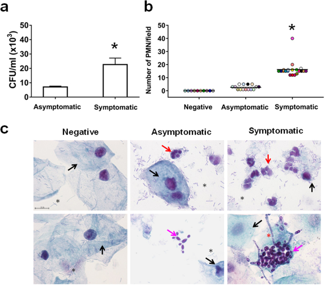
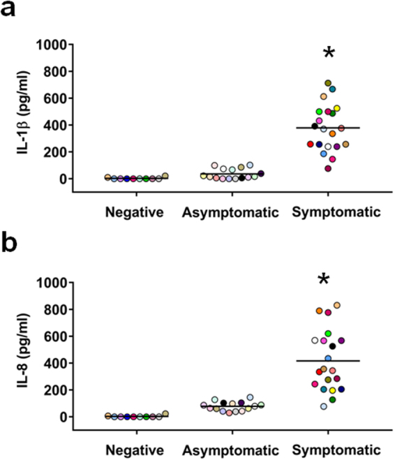
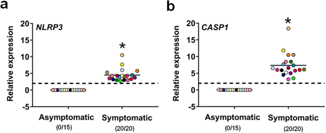
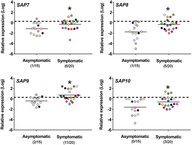
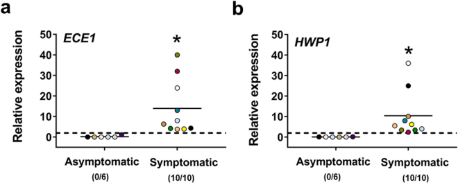
The expression of host inflammatory and Candida albicans putative virulence factors was studied in women with vulvovaginal candidiasis (VVC; twenty) or colonized by the fungus but asymptomatic (carriers; fifteen) or non-colonized asymptomatic (ten subjects). Overexpression of genes encoding NLRP3 and caspase-1 inflammasome components sharply differentiated VVC patients from asymptomatic colonized or non-colonized women. Inflammasome expression was coupled with neutrophils recruitment in the vagina of VVC women and IL-1β and IL-8 production. Both cytokines were present, though to a lower concentration, also in the vaginal fluid of colonized and non-colonized women. Secretory aspartyl proteinases (SAPs) and hyphae associated genes HWP1 and ECE1 were upregulated in VVC but with some differences among infected women. The most overexpressed SAP gene was SAP2, that correlated with neutrophils accumulation. Our data provide clinical evidence that the intracytoplasmic activation of NLRP3 inflammasome complex plays a critical, pathogenesis-relevant role in human VVC.


Conflict of interest statement

The authors declare that they have no competing interests.

Figures

Figure 1
Figure 1
Determination of fungal burden and PMN infiltration in vaginal samples. The CFU in vaginal samples of asymptomatic (n = 5) and symptomatic (n = 5) women were evaluated and the statistical significance was determined with Student’s t test. Data are expressed as mean ± SEM. *p = 0.016 symptomatic vs asymptomatic women (a). Vaginal samples of negative (n = 10, identified by dots with different colors), asymptomatic (n = 15, identified by dots with different colors) and symptomatic (n = 20, identified by dots with different colors) women were examined under light microscope to evaluate the PMN recruitment by morphology. The statistical significance was determined with Mann Whitney U test. *p = 3.07 × 10−10 symptomatic vs asymptomatic women (b). Vaginal samples of negative (n = 10), asymptomatic (n = 15) and symptomatic (n = 20) women were microscopically examined to evaluate the presence of epithelial cells (), PMN (), lactobacilli (*) and C. albicans in yeast () or hyphal form (*) (original magnification x1000, bar = 10 μm). Representative images of each kind of vaginal samples (upper and lower panels) shown are from three different women (c).
Figure 2
Figure 2
IL-1β and IL-8 production in vaginal samples. The supernatants of vaginal samples of negative (n = 10, identified by dots with different colors), asymptomatic (n = 15, identified by dots with different colors) and symptomatic (n = 20, identified by dots with different colors) women were tested for IL-1β (a) and IL-8 (b) production by specific ELISA assays. The results are from triplicates samples of each subjects and the statistical significance was determined with Mann Whitney U test. *p = 2.1 × 10−9 symptomatic vs asymptomatic women for IL-1β. *p = 4.2 × 10−8 symptomatic vs asymptomatic women for IL-8.
Figure 3
Figure 3
Quantitative analysis of NLRP3 and CASP1 gene expression. Vaginal samples of asymptomatic (n = 15, identified by dots with different colors) and symptomatic (n = 20, identified by dots with different colors) women were centrifuged at 3000 rpm for 10 min, then cellular fractions were lysed and total RNA was extracted and retro-transcribed in cDNA. The expression levels of NLRP3 (a) and CASP1 (b) genes in asymptomatic and symptomatic women were calculated by comparative Ct method (2−∆Ct formula) after normalization with GADPH gene. The results are from triplicates samples of each subject and the statistical significance was determined with Mann Whitney U test. The dashed line denotes the cutoff at which the overexpression of NLRP3 and CASP1 genes was defined as >2 times the value of GADPH expression and the number of women expressing genes is indicated in the brackets. *p = 3 × 10−10 symptomatic vs asymptomatic women for NLRP3 and CASP1.
Figure 4
Figure 4
Quantitative analysis of SAP1-6 gene expression. Vaginal samples of asymptomatic (n = 15, identified by dots with different colors) and symptomatic (n = 20, identified by dots with different colors) women were centrifuged at 3000 rpm for 10 min, then cellular fractions were lysed and total RNA was extracted and retro-transcribed in cDNA. The expression levels of SAP1, SAP2, SAP3 (a) and SAP4, SAP5, SAP6 (b) genes in asymptomatic and symptomatic women were calculated by comparative Ct method (2−∆Ct formula) after normalization with ACT1 gene. The results are from triplicates samples of each subject and the statistical significance was determined with Mann Whitney U test. The dashed line denotes the cutoff at which the overexpression of SAP1-6 genes was defined as >2 times the value of ACT1 expression and the number of women expressing genes is indicated in the brackets. *p = 0.01168 symptomatic vs asymptomatic women for SAP1; *p = 5.8 × 10−8 symptomatic vs asymptomatic women for SAP2; *p = 0.04212 symptomatic vs asymptomatic women for SAP3. *p = 0.00014 symptomatic vs asymptomatic women for SAP4; *p = 3.4 × 10−6 symptomatic vs asymptomatic women for SAP5; *p = 0.00029 symptomatic vs asymptomatic women for SAP6.
Figure 5
Figure 5
Quantitative analysis of SAP7-10 gene expression. Vaginal samples of asymptomatic (n = 15, identified by dots with different colors) and symptomatic (n = 20, identified by dots with different colors) women were centrifuged at 3000 rpm for 10 min, then cellular fractions were lysed and total RNA was extracted and retro-transcribed in cDNA. The expression levels of SAP7, SAP8, SAP9 and SAP10 genes in asymptomatic and symptomatic women were calculated by comparative Ct method (2−∆Ct formula) after normalization with ACT1 gene. The results are from triplicates samples of each subject and the statistical significance was determined with Mann Whitney U test. The dashed line denotes the cutoff at which the overexpression of SAP7-10 genes was defined as >2 times the value of ACT1 expression and the number of women expressing genes is indicated in the brackets. *p = 0.00961 symptomatic vs asymptomatic women for SAP7; *p = 0.00457 symptomatic vs asymptomatic women for SAP8; *p = 0.00165 symptomatic vs asymptomatic women for SAP9; *p = 0.02851 symptomatic vs asymptomatic women for SAP10.
Figure 6
Figure 6
Quantitative analysis of ECE1 and HWP1 gene expression. Vaginal samples of asymptomatic (n = 6, identified by dots with different colors) and symptomatic (n = 10, identified by dots with different colors) women were centrifuged at 3000 rpm for 10 min, then cellular fractions were lysed and total RNA was extracted and retro-transcribed in cDNA. The expression levels of ECE1 (a) and HWP1 (b) genes in asymptomatic and symptomatic women were calculated by comparative Ct method (2−∆Ct formula) after normalization with ACT1 gene. The results are from triplicates samples of each subject and the statistical significance was determined with Mann Whitney U test. The dashed line denotes the cutoff at which the overexpression of ECE1 and HWP1 genes was defined as >2 times the value of ACT1 expression and the number of women expressing genes is indicated in the brackets. *p = 0.00012 symptomatic vs asymptomatic women for ECE1 and HWP1.

References

    1. Vecchiarelli A, Pericolini E, Gabrielli E, Pietrella D. New approaches in the development of a vaccine for mucosal candidiasis: progress and challenges. Front Microbiol. 2012;3:294. doi: 10.3389/fmicb.2012.00294. - DOI - PMC - PubMed
    1. Sobel JD. Recurrent vulvovaginal candidiasis. Am J Obstet Gynecol. 2016;214:15–21. doi: 10.1016/j.ajog.2015.06.067. - DOI - PubMed
    1. Vecchiarelli A, Gabrielli E, Pericolini E. Experimental models of vaginal candidiasis and inflammation. Future Microbiol. 2015;10:1265–1268. doi: 10.2217/FMB.15.52. - DOI - PubMed
    1. Cassone A, Sobel JD. Experimental Models of Vaginal Candidiasis and Their Relevance to Human Candidiasis. Infect Immun. 2016;84:1255–1261. doi: 10.1128/IAI.01544-15. - DOI - PMC - PubMed
    1. Bruno, V. M. et al. Transcriptomic analysis of vulvovaginal candidiasis identifies a role for the NLRP3 inflammasome. MBio6, 10.1128/mBio.00182-15 (2015). - PMC - PubMed

Publication types

LinkOut - more resources