. 2017 Dec 19;7(1):17759.
doi: 10.1038/s41598-017-17902-0.

Ubiquitin Ligase Huwe1 Modulates Spermatogenesis by Regulating Spermatogonial Differentiation and Entry into Meiosis

Affiliations

Ubiquitin Ligase Huwe1 Modulates Spermatogenesis by Regulating Spermatogonial Differentiation and Entry into Meiosis

Rohini Bose et al. Sci Rep. .

Abstract

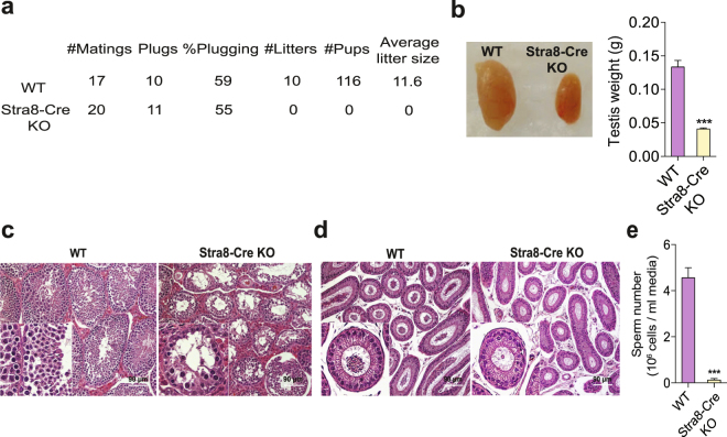
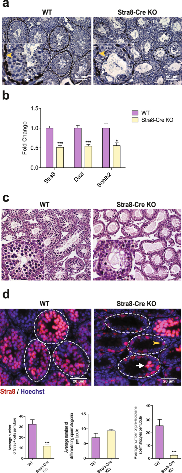
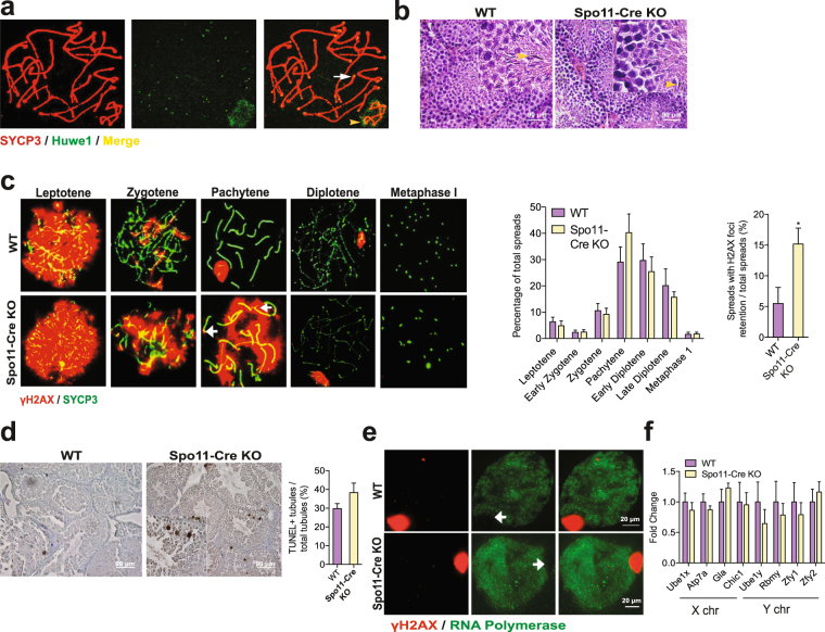
Spermatogenesis consists of a series of highly regulated processes that include mitotic proliferation, meiosis and cellular remodeling. Although alterations in gene expression are well known to modulate spermatogenesis, posttranscriptional mechanisms are less well defined. The ubiquitin proteasome system plays a significant role in protein turnover and may be involved in these posttranscriptional mechanisms. We previously identified ubiquitin ligase Huwe1 in the testis and showed that it can ubiquitinate histones. Since modulation of histones is important at many steps in spermatogenesis, we performed a complete characterization of the functions of Huwe1 in this process by examining the effects of its inactivation in the differentiating spermatogonia, spermatocytes and spermatids. Inactivation of Huwe1 in differentiating spermatogonia led to their depletion and formation of fewer pre-leptotene spermatocytes. The cell degeneration was associated with an accumulation of DNA damage response protein γH2AX, impaired downstream signalling and apoptosis. Inactivation of Huwe1 in spermatocytes indicated that Huwe1 is not essential for meiosis and spermiogenesis, but can result in accumulation of γH2AX. Collectively, these results provide a comprehensive survey of the functions of Huwe1 in spermatogenesis and reveal Huwe1's critical role as a modulator of the DNA damage response pathway in the earliest steps of spermatogonial differentiation.


Conflict of interest statement

The authors declare that they have no competing interests.

Figures

Figure 1
Figure 1
Inactivation of Huwe1 in the differentiating spermatogonia leads to an arrest in spermatogenesis. (a) Inactivation of Huwe1 in the differentiating spermatogonia leads to infertility. WT and Stra8-Cre KO (n = 5) male mice were mated with CD1 females for 4 consecutive weeks. The females were changed every week. Indicated are the frequency of vaginal plugging, number of litters sired and the average litter size. (b)Adult Stra8-Cre KO mice have smaller testes than WT mice. Representative images (left panel) and weights (right panel) of testis from WT (n = 9) and Stra8-Cre KO (n = 10) mice. (c) Arrested spermatogenesis in the Stra8-Cre KO testis. Hematoxylin and eosin stained testicular tissue sections from adult (4 month old) WT and Stra8-Cre KO testes. Scale bar = 90 μm. (d) Few sperm in the Stra8-Cre KO epididymis. Hematoxylin and eosin stained caput (head of the epididymis) tissue sections from adult WT and Stra8-Cre KO. Scale bar = 90 μm. (e) Low sperm number in the Stra8-Cre KO mice. Measurement of sperm number in the cauda (tail of the epididymis) of WT and Stra8-Cre KO (n = 5) mice. Data are shown as mean ± SEM. Student’s t-test. ***p < 0.001.
Figure 2
Figure 2
Huwe1 is a critical regulator of spermatogonial differentiation. (a) Disrupted formation of pre-leptotene spermatocytes in synchronized Stra8-Cre KO testis 48 days post retinoic acid (dpRA) adult testes. Representative immunohistochemical images from WT and Stra8-Cre KO testis sections stained for Stra8. Pre-leptotene spermatocytes are identified based on their Stra8 positivity and location in the seminiferous tubules away from the basement membrane. The yellow arrowhead indicates an A1 differentiating spermatogonia and the white arrow indicates a pre-leptotene spermatocyte. Scale bar = 90 μm. (b) Expression of markers for differentiating spermatogonia is decreased in synchronized Stra8-Cre KO 48 dpRA adult testes. Q-PCR analysis of markers of differentiating spermatogonia (Stra8, Dazl, Sohlh2) in the WT (n = 5) and Stra8-Cre KO (n = 6) testis. (c) Loss of pre-leptotene/leptotene spermatocytes (germ cells located away from the basement membrane) in synchronized Stra8-Cre KO 8 dpRA tubules. Hematoxylin and eosin stained testicular tissue sections from neonatal WT and Stra8-Cre KO mice. Scale bar = 90 μm. (d) Few pre-leptotene spermatocytes in the synchronized Stra8-Cre KO 8 dpRA testis. Representative immunofluorescence images of synchronized WT and Stra8-Cre KO testis stained for Stra8 (red) (top panel). Stra8 + cells at the basement membrane of tubule are A1 differentiating spermatogonia (arrowhead); Stra8 + cells away from the basement membrane are pre-leptotene spermatocytes (arrow). Quantification from the images (bottom panel). WT, n = 5 (135 tubules) Stra8-Cre KO, n = 7 (271 tubules). Scale bar = 20 μm. Data are shown as mean ± SEM. Student’s t-test. ***p < 0.001, *p < 0.05.
Figure 3
Figure 3
Huwe1 is important for entry into meiosis. (a) Inactivation of Huwe1 in the differentiating spermatogonia leads to a defect in proliferation. Representative immunofluorescence images of synchronized 8 dpRA WT and Stra8-Cre KO testis stained for Stra8 (red) and BrdU (green) (top panel). Stra8 + cells at the basement membrane of tubule are A1 differentiating spermatogonia; Stra8 + cells away from the basement membrane are pre-leptotene spermatocytes. The yellow arrowhead indicates an A1 differentiating spermatogonium and the white arrow indicates a pre-leptotene spermatocyte. Quantification from these images (bottom panel) WT, n = 5 (135 tubules), Stra8-Cre KO, n = 7 (271 tubules). Scale bar = 20 μm. (b) Expression of meiotic markers is decreased in the Stra8-Cre KO 8 dpRA testes. Q-PCR analysis of markers of meiosis (Spo11, Smc1b, Sycp1, Sycp3) on WT and Stra8-Cre KO 8 dpRA testes (WT, n = 5, Stra8-Cre KO, n = 6). (c) Disrupted formation of leptotene spermatocytes in Stra8-Cre KO 8 dpRA testes. Representative immunofluorescence images from WT and Stra8-Cre KO testis sections stained for SYCP3 (green). Scale bar = 20 μm. Data are shown as mean ± SEM. Student’s t-test. *p < 0.05.
Figure 4
Figure 4
Inactivation of Huwe1 in the differentiating spermatogonia leads to an activation of DDR and apoptotic cell death. (a) Loss of Huwe1 resulted in elevated proportion of Stra8 + cells showing intense γH2AX foci in unsynchronized 10 dpp testis. Representative immunofluorescence images from unsynchronized 10 dpp WT and Stra8-Cre KO testes stained for γH2AX (green) and Stra8 (red) (left panel). γH2AX+ Stra8- cells in the tubules are germ cells in early meiosis (leptotene/zygotene). The yellow arrowheads indicate Stra8 + differentiating spermatogonia/pre-leptotene spermatocytes and the white arrow indicates a Stra8+ cell that is also γH2AX+ . Quantification of the proportion of Stra8+ cells that are γH2AX + from the images (right panel) WT, n = 4 (89 tubules), KO, n = 4 (128 tubules). Scale bar = 50 μm. (b) Inactivation of Huwe1 leads to the recruitment of pCHK2 to the intense γH2AX foci in unsynchronized 10 dpp testis. Representative images from immunofluorescence staining of 10 dpp WT and Stra8-Cre KO testes stained for γH2AX (green) and pCHK2 (red). Scale bar = 20 μm. (c) Loss of Huwe1 in the differentiating spermatogonia resulted in increased levels of apoptosis in the synchronized 8 dpRA testes. Shown are typical images from TUNEL assay performed on testes sections at 8 dpRA (left panel). The yellow arrowheads indicate apoptotic cells. Scale bar = 90 μm. Quantification from the images (right panel). WT, n = 5 (783 tubules) Stra8-Cre KO, n = 7 (1099 tubules). (d) Loss of Huwe1 resulted in an elevated percentage of cleaved caspase 3 positive tubules in the synchronized 8 dpRA testes. Shown are typical IHC images for cleaved caspase 3 in 8 dpRA WT and Stra8-Cre KO testes (left panel). The yellow arrowhead indicates a cleaved caspase 3 positive cell. Scale bar = 90 μm. Quantification from the images (right panel) WT, n = 4 (269 tubules), KO, n = 4 (286 tubules). Data are shown as mean ± SEM. Student’s t-test. *p < 0.05.
Figure 5
Figure 5
Huwe1 is not essential for completion of the later stages of meiosis. (a) Huwe1 is localized to the sex body and telomeres in pachytene spermatocytes. Representative immunofluorescence image from a chromosome spread prepared from a WT testis at 28 dpp stained for Huwe1 (green) and SYCP3 (red). White arrow points to a Huwe1 + telomere end, yellow arrowhead points to the Huwe1 + sex body. Scale bar = 20 μm. (b) The Spo11-Cre KO animals have normal spermatogenesis. Hematoxylin and eosin stained testicular tissue sections from adult WT and Spo11-Cre KO. The yellow arrowheads indicate spermatids in the lumen of the seminiferous tubules. Scale bar = 90 μm (top panel). (c) The Spo11-Cre KO mice show the retention of γH2AX on the autosomes after homologous recombination. Representative immunofluorescence images of chromosome spreads prepared from WT and Spo11-Cre KO testes at 28 dpp stained for γH2AX (green) and SYCP3 (red) (left panel). White arrow indicates γH2AX foci on an autosome of a pachytene spermatocyte, an abnormality significantly more common in the Spo11-Cre KO testes. Quantification of different cell types from the images and of retention of γH2AX (right panel) WT, n = 4 (402 spreads), Spo11-Cre KO, n = 6 (613 spreads). (d) Spo11-Cre KO of Huwe1 did not lead to increased apoptosis. Shown are typical images from TUNEL assay performed on adult testes sections of WT and Spo11-Cre KO testes (left panel). Scale bar = 90 μm. Quantification from images (right panel) WT, n = 4 (335 tubules), KO, n = 4 (466 tubules). (e) RNA polymerase II remains excluded from the XY body of the Spo11-Cre KO spermatocytes. Representative immunofluorescence images of chromosome spreads prepared from WT and Spo11-Cre KO testes at 28 dpp stained for γH2AX (red) and RNA polymerase II (green) (n = 5). The white arrows indicate the absence of RNA polymerase II stain on the XY body. Scale bar = 20 μm. (f) Normal meiotic sex chromosome inactivation (MSCI) in the Spo11-Cre KO mice. Q-PCR measurements of mRNA levels of selected X-linked and Y-linked genes (n = 5). Data are represented as mean ± SEM. Student’s t-test. *p < 0.05.

References

    1. de Rooij DG. Stem cells in the testis. Int. J. Exp. Pathol. 1998;79:67–80. doi: 10.1046/j.1365-2613.1998.00057.x. - DOI - PMC - PubMed
    1. Griswold MD, Hogarth CA, Bowles J, Koopman P. Initiating meiosis: the case for retinoic acid. Biol. Reprod. 2012;86:35. doi: 10.1095/biolreprod.111.096610. - DOI - PMC - PubMed
    1. Huckins C. The spermatogonial stem cell population in adult rats. I. Their morphology, proliferation and maturation. Anat. Rec. 1971;169:533–557. doi: 10.1002/ar.1091690306. - DOI - PubMed
    1. Oakberg EF. Spermatogonial stem-cell renewal in the mouse. Anat. Rec. 1971;169:515–531. doi: 10.1002/ar.1091690305. - DOI - PubMed
    1. Baudat F, Manova K, Yuen JP, Jasin M, Keeney S. Chromosome synapsis defects and sexually dimorphic meiotic progression in mice lacking Spo11. Mol. Cell. 2000;6:989–998. doi: 10.1016/S1097-2765(00)00098-8. - DOI - PubMed

Publication types

MeSH terms

Grants and funding

LinkOut - more resources